Macular Choroidal Thickness and Volume Measured by Swept-source Optical Coherence Tomography in Healthy Korean Children
- Affiliations
-
- 1Department of Ophthalmology, Hanyang University College of Medicine, Seoul, Korea. brlee@hanyang.ac.kr
- 2102 Replacement Depot, Republic of Korea Army, Chuncheon, Korea.
- KMID: 2363888
- DOI: http://doi.org/10.3341/kjo.2016.30.1.32
Abstract
- PURPOSE
To evaluate the thickness and volume of the choroid in healthy Korean children using swept-source optical coherence tomography.
METHODS
We examined 80 eyes of 40 healthy children and teenagers (<18 years) using swept-source optical coherence tomography with a tunable long-wavelength laser source. A volumetric macular scan protocol using the Early Treatment Diabetic Retinopathy Study grid was used to construct a choroidal thickness map. We also examined 44 eyes of 35 healthy adult volunteers (> or =18 years) and compared adult measurements with the findings in children.
RESULTS
The mean age of the children and teenagers was 9.47 +/- 3.80 (4 to 17) vs. 55.04 +/- 12.63 years (36 to 70 years) in the adult group (p < 0.001, Student's t-test). Regarding the Early Treatment Diabetic Retinopathy Study subfields, the inner temporal subfield was the thickest (247.96 microm). The inner and outer nasal choroid were thinner (p = 0.004, p = 0.002, respectively) than the surrounding areas. The mean choroidal volumes of the inner and outer nasal areas were smaller (p = 0.004, p = 0.003, respectively) than those of all the other areas in each circle. Among the nine subfields, all areas in the children, except the outer nasal subfield, were thicker than those in adults (p < 0.05). Regression analysis showed that age, axial length, and refractive error correlated with subfoveal choroidal thickness (p < 0.05).
CONCLUSIONS
Overall macular choroidal thickness and volume in children and teenagers were significantly greater than in adults. The nasal choroid was significantly thinner than the surrounding areas. The pediatric subfoveal choroid is prone to thinning with increasing age, axial length, and refractive error. These differences should be considered when choroidal thickness is evaluated in children with chorioretinal diseases.
Keyword
MeSH Terms
Figure
-
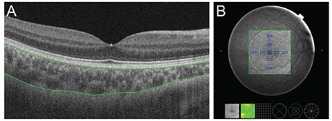
Fig. 1 Choroidal thickness map obtained using swept-source optical coherence tomography in a healthy 9-year-old girl. The 3-dimensional (D) volumetric macular scan protocol with 512 A-scans × 256 B-scans was used to obtain 3D imaging data of a 6 × 6 mm area. (A) Autosegmentation of the chorioscleral border in the B-scan image. (B) Choroidal thickness map of a 6 × 6 mm area centered on the fovea obtained from analysis of the B-scan images in the 3D data set. Mean choroidal thickness for each sector was obtained by applying the Early Treatment Diabetic Retinopathy Study grid to the map.
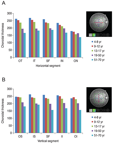
Fig. 2 Pediatric and adult choroidal thinning with increasing age in each (A) horizontal and (B) vertical segment of the Early Treatment Diabetic Retinopathy Study grid. OT = outer temporal; IT = inner temporal; SF = subfoveal; IN = inner nasal; ON = outer nasal; OS = outer superior; IS = inner superior; II = inner inferior; OI = outer inferior.
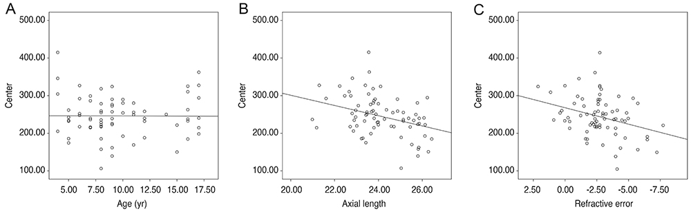
Fig. 3 Scatterplots show the relationship between central choroidal thickness and (A) age, (B) axial length, and (C) refractive error. As age and axial length increased, central choroidal thickness (A,B) decreased. In contrast, as the spherical equivalent increased, central choroidal thickness (C) tended to increase.
Reference
-
1. Harris A, Bingaman D, Ciulla T, Martin B. Retinal and choroidal blood flow in health and disease. In : Ryan SJ, Hinton DR, Schachat AP, Wilkinson P, editors. Retina. 4th ed. Philadelphia: Elsevier;2006. p. 83–102.2. Kim KH, Kim DG. The relationship among refractive power, axial length and choroidal thickness measured by SD-OCT in myopia. J Korean Ophthalmol Soc. 2012; 53:626–631.3. Ikuno Y, Kawaguchi K, Nouchi T, Yasuno Y. Choroidal thickness in healthy Japanese subjects. Invest Ophthalmol Vis Sci. 2010; 51:2173–2176.4. Kim SW, Oh J, Kwon SS, et al. Comparison of choroidal thickness among patients with healthy eyes, early age-related maculopathy, neovascular age-related macular degeneration, central serous chorioretinopathy, and polypoidal choroidal vasculopathy. Retina. 2011; 31:1904–1911.5. Cho JH, Bae SH, Han JR, et al. Comparison of choroidal thickness in eyes with central serous chorioretinopathy, asymptomatic fellow eyes and normal eyes. J Korean Ophthalmol Soc. 2012; 53:87–93.6. Chung SE, Kang SW, Lee JH, Kim YT. Choroidal thickness in polypoidal choroidal vasculopathy and exudative age-related macular degeneration. Ophthalmology. 2011; 118:840–845.7. Manjunath V, Goren J, Fujimoto JG, Duker JS. Analysis of choroidal thickness in age-related macular degeneration using spectral-domain optical coherence tomography. Am J Ophthalmol. 2011; 152:663–668.8. Spaide RF, Koizumi H, Pozzoni MC. Enhanced depth imaging spectral-domain optical coherence tomography. Am J Ophthalmol. 2008; 146:496–500.9. Margolis R, Spaide RF. A pilot study of enhanced depth imaging optical coherence tomography of the choroid in normal eyes. Am J Ophthalmol. 2009; 147:811–815.10. Spaide RF, Akiba M, Ohno-Matsui K. Evaluation of peripapillary intrachoroidal cavitation with swept source and enhanced depth imaging optical coherence tomography. Retina. 2012; 32:1037–1044.11. Nagasawa T, Mitamura Y, Katome T, et al. Swept-source optical coherence tomographic findings in morning glory syndrome. Retina. 2014; 34:206–208.12. Hirata M, Tsujikawa A, Matsumoto A, et al. Macular choroidal thickness and volume in normal subjects measured by swept-source optical coherence tomography. Invest Ophthalmol Vis Sci. 2011; 52:4971–4978.13. Ruiz-Moreno JM, Flores-Moreno I, Lugo F, et al. Macular choroidal thickness in normal pediatric population measured by swept-source optical coherence tomography. Invest Ophthalmol Vis Sci. 2013; 54:353–359.14. Read SA, Collins MJ, Vincent SJ, Alonso-Caneiro D. Choroidal thickness in childhood. Invest Ophthalmol Vis Sci. 2013; 54:3586–3593.15. Nagasawa T, Mitamura Y, Katome T, et al. Macular choroidal thickness and volume in healthy pediatric individuals measured by swept-source optical coherence tomography. Invest Ophthalmol Vis Sci. 2013; 54:7068–7074.16. Early Treatment Diabetic Retinopathy Study design and baseline patient characteristics. ETDRS report number 7. Ophthalmology. 1991; 98:741–756.17. Brown JS, Flitcroft DI, Ying GS, et al. In vivo human choroidal thickness measurements: evidence for diurnal fluctuations. Invest Ophthalmol Vis Sci. 2009; 50:5–12.18. Tan CS, Ouyang Y, Ruiz H, Sadda SR. Diurnal variation of choroidal thickness in normal, healthy subjects measured by spectral domain optical coherence tomography. Invest Ophthalmol Vis Sci. 2012; 53:261–266.19. Kim EJ, Kim JH, Koo SH, et al. Choroidal thickness changes according to the refractive errors and axial length in Korean myopia patients. J Korean Ophthalmol Soc. 2012; 53:1814–1822.20. Li XQ, Larsen M, Munch IC. Subfoveal choroidal thickness in relation to sex and axial length in 93 Danish university students. Invest Ophthalmol Vis Sci. 2011; 52:8438–8441.21. Kim JH, Kim JS, Lee KW, Lee JH. The posterior choroidal profiles measured by spectral domain optical coherence tomography in healthy Korean children. J Korean Ophthalmol Soc. 2013; 54:1708–1714.22. Shin JW, Shin YU, Lee BR. Choroidal thickness and volume mapping by a six radial scan protocol on spectral-domain optical coherence tomography. Ophthalmology. 2012; 119:1017–1023.
- Full Text Links
-
- Actions
-
Cited
- CITED
-
- Close
- Share
-
- Similar articles
-
- Macular Choroidal Thickness and Volume Measured by Swept-source Optical Coherence Tomography in Healthy Korean Children
- Choroidal Thickness at the Outside of Fovea in Diabetic Retinopathy Using Spectral-Domain Optical Coherence Tomography
- Utility of the Swept Source Optical Coherence Tomography for Measurements of Central Corneal Thickness
- Diagnostic Ability of Macular Ganglion Cell Layer Measurements in Glaucoma Using Swept Source Optical Coherence Tomography
- The Relationship among Refractive Power, Axial Length and Choroidal Thickness Measured by SD-OCT in Myopia
