Argon Green Laser for Valsalva Retinopathy Treatment and Long-term Follow-up of the Internal Limiting Membrane Changes in Optical Coherence Tomography
- Affiliations
-
- 1Ulucanlar Eye Training and Research Hospital, Ankara, Turkey.
- 2Department of Ophthalmology, Faculty of Medicine, Hitit University, Corum, Turkey. cagataycaglar@hitit.edu.tr
- 3Ulucanlar Eye Training and Research Hospital, Ankara, Turkey.
- KMID: 2363854
- DOI: http://doi.org/10.3341/kjo.2015.29.6.437
Abstract
- No abstract available.
MeSH Terms
Figure
-
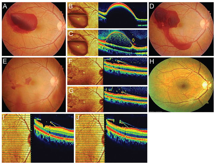
Fig. 1 (A) Fundus photograph of the right eye showed a huge sub-internal limiting membrane (ILM) hemorrhage in the temporal macular area. (B) Spectral domain optic coherence tomography (OCT) revealed a dome-shaped hypo-reflective area, consistent with blood beneath a hyper-reflective band at the macula. (C) Spectral domain OCT reveals two distinct membranes: the more apparent and reflective one was identified as ILM (red arrow), and the other slight and patched membrane was identified as posterior hyaloid (yellow arrow). (D) The hemorrhage was drained inferiorly after the argon laser procedure. (E) Fundus photograph reveals strong resolution of the hemorrhage after three weeks. (F,G) OCT reveals laser perforation points along the ILM and hyaloid after three weeks. (H) There was no hemorrhage in fundus photograph after two years. (I) Pseudo-hole and no reattachment of ILM are observed in the temporal macular area in the last OCT. (J) OCT reveals that the detached ILM was attached to the posterior hyaloid and formation of no epiretinal membrane after two years.
Reference
-
1. Schuman JS, Puliafito CA, Fujimoto JG, editors. Optical coherence tomography of ocular diseases. 2nd ed. Thorofare: Slack;2004. p. 1–698.2. Meyer CH, Mennel S, Rodrigues EB, Schmidt JC. Is the location of valsalva hemorrhages submembranous or subhyaloidal? Am J Ophthalmol. 2006; 141:231.3. Shukla D, Naresh KB, Kim R. Optical coherence tomography findings in valsalva retinopathy. Am J Ophthalmol. 2005; 140:134–136.4. Goel N, Kumar V, Seth A, et al. Spectral-domain optical coherence tomography following Nd:YAG lasermembranotomy in valsalva retinopathy. Ophthalmic Surg Lasers Imaging. 2011; 42:222–228.
- Full Text Links
-
- Actions
-
Cited
- CITED
-
- Close
- Share
-
- Similar articles
-
- A Case of Green Laser Pointer-induced Atypical Impending Macular Hole Treated with Vitrectomy in a Pediatric Patient
- Diode Laser Panretinal Photocoagulation in Diabetic Retinopathy
- Macular Hole Formation after Pars Plana Vitrectomy for the Treatment of Valsalva Retinopathy: A Case Report
- A Case of Maculopathy from Handheld Green Laser Pointer
- Change in Subfoveal Choroidal Thickness after Argon Laser Panretinal Photocoagulation
