Korean J Ophthalmol.
2015 Dec;29(6):418-423. 10.3341/kjo.2015.29.6.418.
Differences between Non-arteritic Anterior Ischemic Optic Neuropathy and Open Angle Glaucoma with Altitudinal Visual Field Defect
- Affiliations
-
- 1Department of Ophthalmology, Kim's Eye Hospital, Seoul, Korea. ungsookim@kimeye.com
- 2Department of Ophthalmology, Konyang University College of Medicine, Daejeon, Korea.
- KMID: 2363850
- DOI: http://doi.org/10.3341/kjo.2015.29.6.418
Abstract
- PURPOSE
To investigate the differences in retinal nerve fiber layer (RNFL) change and optic nerve head parameters between non-arteritic anterior ischemic optic neuropathy (NAION) and open angle glaucoma (OAG) with altitudinal visual field defect.
METHODS
Seventeen NAION patients and 26 OAG patients were enrolled prospectively. The standard visual field indices (mean deviation, pattern standard deviation) were obtained from the Humphrey visual field test and differences between the two groups were analyzed. Cirrus HD-OCT parameters were used, including optic disc head analysis, average RNFL thickness, and RNFL thickness of each quadrant.
RESULTS
The mean deviation and pattern standard deviation were not significantly different between the groups. In the affected eye, although the disc area was similar between the two groups (2.00 +/- 0.32 and 1.99 +/- 0.33 mm2, p = 0.586), the rim area of the OAG group was smaller than that of the NAION group (1.26 +/- 0.56 and 0.61 +/- 0.15 mm2, respectively, p < 0.001). RNFL asymmetry was not different between the two groups (p = 0.265), but the inferior RNFL thickness of both the affected and unaffected eyes were less in the OAG group than in the NAION group. In the analysis of optic disc morphology, both affected and unaffected eyes showed significant differences between two groups.
CONCLUSIONS
To differentiate NAION from OAG in eyes with altitudinal visual field defects, optic disc head analysis of not only the affected eye, but also the unaffected eye, by using spectral domain optical coherence tomography may be helpful.
MeSH Terms
-
Aged
Arteritis/diagnosis
Diagnosis, Differential
Female
Glaucoma, Open-Angle/*diagnosis
Humans
Male
Middle Aged
Nerve Fibers/*pathology
Optic Disk/*pathology
Optic Neuropathy, Ischemic/*diagnosis
Prospective Studies
Retinal Ganglion Cells/*pathology
Tomography, Optical Coherence
Vision Disorders/*diagnosis
Visual Field Tests
*Visual Fields
Figure
-
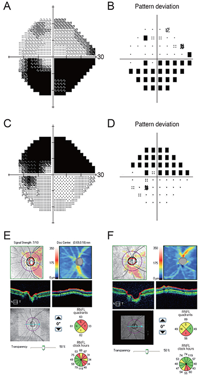
Fig. 1 The results of visual field test (A-D) and spectral domain optical coherence (E,F). Ischemic optic neuropathy (A,B,E) and open angle glaucoma (C,D,F). RNFL = retinal nerve fiber layer.

Fig. 2 Retinal nerve fiber layer thickness analysis using spectral domain optical coherence tomography in the unaffected eye. Inferior retinal nerve fiber layer thickness showed a significant difference between the two groups (p < 0.012, Mann-Whitney test). AION = anterior ischemic optic neuropathy. *p < 0.001.
Reference
-
1. Sethi HS, Lam BL, Romano JG. Reversible prolonged bilateral inferior altitudinal visual field defects associated with migraine. J Neuroophthalmol. 2012; 32:252–255.2. Shapey J, Danesh-Meyer HV, Kaye AH. Suprasellar meningioma presenting with an altitudinal field defect. J Clin Neurosci. 2012; 19:155–158.3. Hayreh SS, Podhajsky PA, Zimmerman MB. Branch retinal artery occlusion: natural history of visual outcome. Ophthalmology. 2009; 116:1188–1194.e4.4. Vuori ML, Mantyjarvi M. Tilted disc syndrome may mimic false visual field deterioration. Acta Ophthalmol. 2008; 86:622–625.5. Hayreh SS, Zimmerman B. Visual field abnormalities in nonarteritic anterior ischemic optic neuropathy: their pattern and prevalence at initial examination. Arch Ophthalmol. 2005; 123:1554–1562.6. Fang JP, Donahue SP, Lin RH. Global visual field involvement in acute unilateral optic neuritis. Am J Ophthalmol. 1999; 128:554–565.7. Suh MH, Kim SH, Park KH, et al. Comparison of the correlations between optic disc rim area and retinal nerve fiber layer thickness in glaucoma and nonarteritic anterior ischemic optic neuropathy. Am J Ophthalmol. 2011; 151:277–286.e1.8. Danesh-Meyer HV, Boland MV, Savino PJ, et al. Optic disc morphology in open-angle glaucoma compared with anterior ischemic optic neuropathies. Invest Ophthalmol Vis Sci. 2010; 51:2003–2010.9. Seymenoglu G, Baser E, Ozturk B. Comparison of spectral-domain optical coherence tomography and Heidelberg retina tomograph III optic nerve head parameters in glaucoma. Ophthalmologica. 2013; 229:101–105.10. Kumar V, Ramanathan US, Mushtaq B, Shah P. Artefactual uniocular altitudinal visual field defect. Br J Ophthalmol. 2002; 86:1442–1443.11. Horowitz J, Fishelzon-Arev T, Rath EZ, et al. Comparison of optic nerve head topography findings in eyes with non-arteritic anterior ischemic optic neuropathy and eyes with glaucoma. Graefes Arch Clin Exp Ophthalmol. 2010; 248:845–851.12. Anton A, Moreno-Montanes J, Blazquez F, et al. Usefulness of optical coherence tomography parameters of the optic disc and the retinal nerve fiber layer to differentiate glaucomatous, ocular hypertensive, and normal eyes. J Glaucoma. 2007; 16:1–8.13. Danesh-Meyer HV, Savino PJ, Sergott RC. The prevalence of cupping in end-stage arteritic and nonarteritic anterior ischemic optic neuropathy. Ophthalmology. 2001; 108:593–598.14. Saito H, Tomidokoro A, Tomita G, et al. Optic disc and peripapillary morphology in unilateral nonarteritic anterior ischemic optic neuropathy and age- and refraction-matched normals. Ophthalmology. 2008; 115:1585–1590.15. Lisboa R, Leite MT, Zangwill LM, et al. Diagnosing preperimetric glaucoma with spectral domain optical coherence tomography. Ophthalmology. 2012; 119:2261–2269.
- Full Text Links
-
- Actions
-
Cited
- CITED
-
- Close
- Share
-
- Similar articles
-
- A Case of Non-Arteritic Anterior Ischemic Optic Neuropathy after Bilateral Selective Neck Dissection
- Temporal Arteritis with Diagnostic Brain Magnetic Resonance Imaging
- Monocular Superior Altitudinal Field defect due to Supraclinoid Internal Carotid Artery Aneurysm
- A Case of Anterior Ischemic Optic Neuropethy
- A Case of Prolonged Bilateral Inferior Altitudinal Visual Field Defect in a Young Migraineur
