A Case of Congenital Simple Hamartoma of the Retinal Pigment Epithelium and Coats' Disease in the Same Eye
- Affiliations
-
- 1Department of Ophthalmology, Kyungpook National University School of Medicine, Daegu, Korea. jps11@hanmail.net
- KMID: 2363774
- DOI: http://doi.org/10.3341/kjo.2015.29.4.282
Abstract
- No abstract available.
MeSH Terms
Figure
-
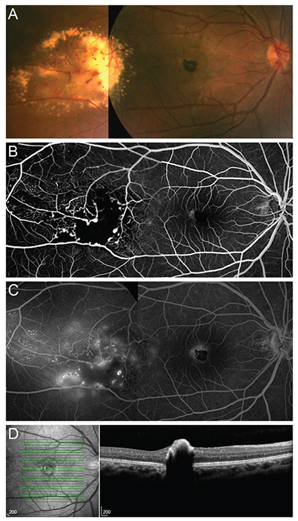
Fig. 1 (A) Fundus photograph at initial examination of the right eye shows a small, well-circumscribed, darkly pigmented mass involving the fovea. Telangiectasia of retinal vessels and yellowish-white exudation are also seen in the temporal midperipheral retina. (B) Early phase of fluorescein angiography shows blockage of fluorescence in the fovea due to a mass lesion and a halo of hyperfluorescence around the foveal mass lesion. In the temporal midperipheral retina, aneurysmal dilatation of retinal vessels and hypofluorescence due to exudation and capillary drop-out are seen. (C) Late phase of fluorescein angiography shows subtle leakage around the foveal mass lesion and vascular leakage in the temporal midperipheral retina. (D) Spectral domain optical coherence tomography of the right eye shows a highly reflective, well-defined retinal elevation with complete optical shadowing of the fovea.
Reference
-
1. Shields CL, Shields JA, Marr BP, et al. Congenital simple hamartoma of the retinal pigment epithelium: a study of five cases. Ophthalmology. 2003; 110:1005–1011.2. Shields JA, Shields CL, Honavar SG, et al. Classification and management of Coats disease: the 2000 Proctor Lecture. Am J Ophthalmol. 2001; 131:572–583.3. Lopez JM, Guerrero P. Congenital simple hamartoma of the retinal pigment epithelium: optical coherence tomography and angiography features. Retina. 2006; 26:704–706.4. Khurana RN, Samuel MA, Murphree AL, et al. Subfoveal nodule in Coats' disease. Clin Experiment Ophthalmol. 2005; 33:301–302.5. Jumper JM, Pomerleau D, McDonald HR, et al. Macular fibrosis in Coats disease. Retina. 2010; 30:4 Suppl. S9–S14.
- Full Text Links
-
- Actions
-
Cited
- CITED
-
- Close
- Share
-
- Similar articles
-
- Ultrastructural Studies of Retina and Proliferative Membranes in Coats' Disease Complicated by Proliferative Vitreoretinopathy
- Growth Patterns of Human Retinal Pigment Epithelium in Vitro
- A Case of Retinoblastoma and Coats' Disease in the Same eye: A Clinicopathologic Report
- Atypical Congenital Hypertrophy of the Retinal Pigment Epithelium in Gardner's Syndrome
- Spectral-domain Optical Coherence Tomography of Combined Hamartoma of the Retina and Retinal Pigment Epithelium in Neurofibromatosis
