Korean J Ophthalmol.
2015 Aug;29(4):220-225. 10.3341/kjo.2015.29.4.220.
Evaluation of Anterior Segment Parameters in Obesity
- Affiliations
-
- 1Department of Ophthalmology, Suleyman Demirel University Medical School, Isparta, Turkey. dralimesefer@hotmail.com
- 2Department of Ophthalmology, Recep Tayyip Erdogan University Medical School, Rize, Turkey.
- 3Department of Ophthalmology, Sorgun State Hospital, Yozgat, Turkey.
- 4Department of Ophthalmology, Gazipasa State Hospital, Antalya, Turkey.
- KMID: 2363765
- DOI: http://doi.org/10.3341/kjo.2015.29.4.220
Abstract
- PURPOSE
To investigate anterior segment parameters in obese patients in comparison to healthy individuals.
METHODS
Thirty-four obese subjects and 34 age-sex-matched healthy subjects were enrolled in this prospective cross-sectional study. Ophthalmological examinations including intraocular pressure (IOP), central corneal thickness (CCT), anterior chamber depth (ACD), anterior chamber volume (ACV), anterior chamber angle (ACA), and axial length (AL) measurements were performed on each subject. Height and weight of all subjects were recorded and body mass index (BMI) was calculated.
RESULTS
IOP was significantly higher in the obese group (p = 0.003). The mean ACD in obese subjects was significantly lower than that in control subjects (p = 0.036). AL, ACV, ACA and CCT were not significantly different between the groups. There was a positive correlation between BMI and IOP (r = 0.404, p < 0.001). ACD and ACA were negatively correlated with BMI.
CONCLUSIONS
IOP was significantly higher and ACD was significantly lower in obese subjects. AL, ACV, ACA and CCT were not significantly different between the groups. The impact of obesity on anterior chamber parameters should be further investigated.
Keyword
MeSH Terms
Figure
-

Fig. 1 Scatter plot between anterior chamber angle (ACA) and anterior chamber depth (ACD) measurements.
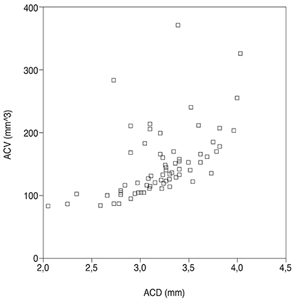
Fig. 2 Scatter plot between anterior chamber volume (ACV) and anterior chamber depth (ACD) measurements.
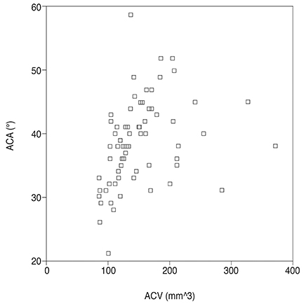
Fig. 3 Scatter plot between anterior chamber angle (ACA) and anterior chamber volume (ACV) measurements.
Reference
-
1. World Health Organization. Controlling the global obesity epidemic. Geneva: World Health Organization;2002. p. 60.2. Eckel RH, Krauss RM. AHA Nutrition Committee. American Heart Association call to action: obesity as a major risk factor for coronary heart disease. Circulation. 1998; 97:2099–2100.3. Grundy SM. Metabolic complications of obesity. Endocrine. 2000; 13:155–165.4. Lawrence VJ, Kopelman PG. Medical consequences of obesity. Clin Dermatol. 2004; 22:296–302.5. Leske MC, Wu SY, Hennis A, et al. Diabetes, hypertension, and central obesity as cataract risk factors in a black population: the Barbados Eye Study. Ophthalmology. 1999; 106:35–41.6. Kuang TM, Tsai SY, Hsu WM, et al. Body mass index and age-related cataract: the Shihpai Eye Study. Arch Ophthalmol. 2005; 123:1109–1114.7. Leske MC, Connell AM, Wu SY, et al. Risk factors for open-angle glaucoma: the Barbados Eye Study. Arch Ophthalmol. 1995; 113:918–924.8. Cheung N, Wong TY. Obesity and eye diseases. Surv Ophthalmol. 2007; 52:180–195.9. Van Leiden HA, Dekker JM, Moll AC, et al. Blood pressure, lipids, and obesity are associated with retinopathy: the hoorn study. Diabetes Care. 2002; 25:1320–1325.10. Seddon JM, Cote J, Rosner B. Progression of age-related macular degeneration: association with dietary fat, transunsaturated fat, nuts, and fish intake. Arch Ophthalmol. 2003; 121:1728–1737.11. Maralani HG, Tai BC, Wong TY, et al. Metabolic syndrome and risk of age-related macular degeneration. Retina. 2015; 35:459–466.12. Nemeth J, Fekete O, Pesztenlehrer N. Optical and ultrasound measurement of axial length and anterior chamber depth for intraocular lens power calculation. J Cataract Refract Surg. 2003; 29:85–88.13. Abolbashari F, Mohidin N, Ahmadi Hosseini SM, et al. Anterior segment characteristics of keratoconus eyes in a sample of Asian population. Cont Lens Anterior Eye. 2013; 36:191–195.14. Lee Y, Sung KR, Na JH, Sun JH. Dynamic changes in anterior segment (AS) parameters in eyes with primary angle closure (PAC) and PAC glaucoma and open-angle eyes assessed using AS optical coherence tomography. Invest Ophthalmol Vis Sci. 2012; 53:693–697.15. Malyugin BE, Shpak AA, Pokrovskiy DF. Accommodative changes in anterior chamber depth in patients with high myopia. J Cataract Refract Surg. 2012; 38:1403–1407.16. Tzamaloukas AH, Murata GH, Hoffman RM, et al. Classification of the degree of obesity by body mass index or by deviation from ideal weight. JPEN J Parenter Enteral Nutr. 2003; 27:340–348.17. Wells JC, Victora CG. Indices of whole-body and central adiposity for evaluating the metabolic load of obesity. Int J Obes (Lond). 2005; 29:483–489.18. Gesta S, Tseng YH, Kahn CR. Developmental origin of fat: tracking obesity to its source. Cell. 2007; 131:242–256.19. Stojanov O, Stokic E, Sveljo O, Naumovic N. The influence of retrobulbar adipose tissue volume upon intraocular pressure in obesity. Vojnosanit Pregl. 2013; 70:469–476.20. Otto AJ, Koornneef L, Mourits MP, et al. Retrobulbar pressures measured during surgical decompression of the orbit. Br J Ophthalmol. 1996; 80:1042–1045.21. Nassr MA, Morris CL, Netland PA, Karcioglu ZA. Intraocular pressure change in orbital disease. Surv Ophthalmol. 2009; 54:519–544.22. Mori K, Ando F, Nomura H, et al. Relationship between intraocular pressure and obesity in Japan. Int J Epidemiol. 2000; 29:661–666.23. Zafra Perez JJ, Villegas Perez MP, Canteras Jordana M, Miralles De Imperial J. Intraocular pressure and prevalence of occult glaucoma in a village of Murcia. Arch Soc Esp Oftalmol. 2000; 75:171–178.24. Jang HD, Kim DH, Han K, et al. Relationship between intraocular pressure and parameters of obesity in Korean adults: the 2008-2010 Korea National Health and Nutrition Examination Survey. Curr Eye Res. 2014; 11. 07. DOI: 10.3109/02713683.2014.975367.25. Newman-Casey PA, Talwar N, Nan B, et al. The relationship between components of metabolic syndrome and open-angle glaucoma. Ophthalmology. 2011; 118:1318–1326.26. Lavanya R, Wong TY, Friedman DS, et al. Determinants of angle closure in older Singaporeans. Arch Ophthalmol. 2008; 126:686–691.27. Lowe RF. Aetiology of the anatomical basis for primary angle-closure glaucoma. Biometrical comparisons between normal eyes and eyes with primary angle-closure glaucoma. Br J Ophthalmol. 1970; 54:161–169.28. Halpern DL, Grosskreutz CL. Glaucomatous optic neuropathy: mechanisms of disease. Ophthalmol Clin North Am. 2002; 15:61–68.29. Saw SM, Chua WH, Hong CY, et al. Height and its relationship to refraction and biometry parameters in Singapore Chinese children. Invest Ophthalmol Vis Sci. 2002; 43:1408–1413.30. Wong TY, Foster PJ, Johnson GJ, et al. The relationship between ocular dimensions and refraction with adult stature: the Tanjong Pagar Survey. Invest Ophthalmol Vis Sci. 2001; 42:1237–1242.31. Praveen MR, Vasavada AR, Shah SK, et al. Lens thickness of Indian eyes: impact of isolated lens opacity, age, axial length, and inf luence on anterior chamber depth. Eye (Lond). 2009; 23:1542–1548.32. Sahinoglu-Keskek N, Keskek SO, Cevher S, et al. Metabolic syndrome as a risk factor for elevated intraocular pressure. Pak J Med Sci. 2014; 30:477–482.33. Su DH, Wong TY, Foster PJ, et al. Central corneal thickness and its associations with ocular and systemic factors: the Singapore Malay Eye Study. Am J Ophthalmol. 2009; 147:709–716.e1.34. Nishitsuka K, Kawasaki R, Kanno M, et al. Determinants and risk factors for central corneal thickness in Japanese persons: the Funagata Study. Ophthalmic Epidemiol. 2011; 18:244–249.35. Yin G, Wang YX, Zheng ZY, et al. Ocular axial length and its associations in Chinese: the Beijing Eye Study. PLoS One. 2012; 7:e43172.
- Full Text Links
-
- Actions
-
Cited
- CITED
-
- Close
- Share
-
- Similar articles
-
- Comparision of Anterior Segment Parameters in Angle-Closure Glaucoma Using Scheimpflug Camera
- Ultrasound Biomicroscopy (UBM plus, model P45, Paradigm(R)): Intraobserver Reproducibility and Agreement of Measurements
- Measurement of Anterior Segment Using Visante OCT in Koreans
- Differences between the Results Assessed by Slit Lamp Examination and Anterior Segment Photography in Terms of Cataract Grading
- Aneurysms of the Proximal Anterior Cerebral Artery
