Korean J Ophthalmol.
2015 Jun;29(3):155-159. 10.3341/kjo.2015.29.3.155.
Demographic Features of Idiopathic Macular Telangiectasia in Korean Patients
- Affiliations
-
- 1Department of Ophthalmology, Dongguk University Ilsan Hospital, Goyang, Korea. blueretinaoh@gmail.com
- 2Department of Ophthalmology, Korea University College of Medicine, Seoul, Korea.
- KMID: 2363752
- DOI: http://doi.org/10.3341/kjo.2015.29.3.155
Abstract
- PURPOSE
To investigate the clinical and demographic features of idiopathic macular telangiectasia (MacTel) in Korean patients since the introduction of spectral domain optical coherence tomography (SD-OCT).
METHODS
We reviewed medical records of patients who were diagnosed with MacTel from 2009 to 2013. All patients underwent fluorescein angiography and SD-OCT and were classified as type 1 or type 2 according to the classification system proposed by Yannuzzi.
RESULTS
Over a period of 5 years, 4 (18.2%) patients were diagnosed with type 1 MacTel and 18 (81.8%) patients were diagnosed with type 2 MacTel. All patients with type1 MacTel were male, and their mean age was 51 +/- 8.6 years. Among patients with type 2 MacTel, 3 (16.7%) were male, 15 (83.3%) were female, and the mean age was 60 +/- 13.6 years. Whereas all type 1 MacTel patients had either metamorphopsia or mild scotoma, of the 18 patients with type 2 MacTel, only 4 (22.2%) had those symptoms, 10 (55.6%) complained of only mild visual impairment, and the other 4 (22.2%) had no symptoms. Intraretinal cystoid spaces were observed in 26 (72.2%) of 36 eyes with type 2 MacTel by SD-OCT. These cystoid spaces had irregular boundaries and did not correspond to angiographic leakages.
CONCLUSIONS
Type 2 MacTel was most common in the present study. The wider availability of SD-OCT may have contributed to the diagnosis of type 2 MacTel. Type 2 MacTel may be more prevalent than type 1 in Koreans, which corresponds to the results of Western countries.
Keyword
MeSH Terms
Figure
-

Fig. 1 Type 1 idiopathic macular telangiectasia. (A) Fundus photograph shows multiple microaneurysms and retinal edema with hard exudates. (B) Late phase of fluorescein angiograph shows telangiectasis and multiple microaneurysms. (C) Spectral domain optical coherence tomography image shows retinal thickening and cystoid spaces.
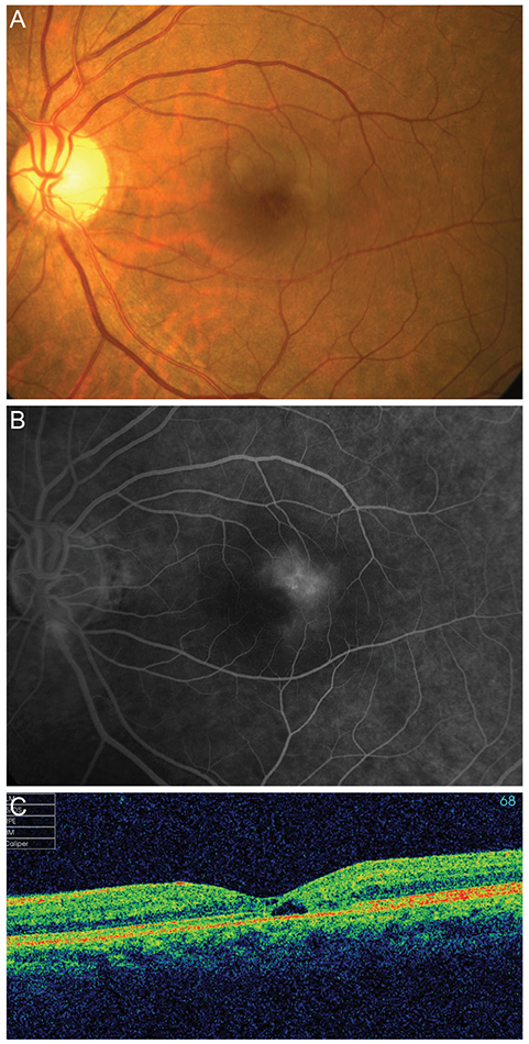
Fig. 2 Type 2 idiopathic macular telangiectasia. (A) Fundus photograph shows slight graying of the perifoveolar retina. (B) Late phase of fluorescein angiograph shows hyperfluorescence temporal to the foveola. (C) Spectral domain optical coherence tomography image shows cystoid spaces without retinal thickening.
Reference
-
1. Engelbert M, Chew EY, Yannuzzi LA. Macular telangiectasia. In : Ryan SJ, editor. Retina. 5th ed. London: Saunders/Elsevier;2013. p. 1050–1057.2. Gass JD, Oyakawa RT. Idiopathic juxtafoveolar retinal telangiectasis. Arch Ophthalmol. 1982; 100:769–780.3. Gass JD, Blodi BA. Idiopathic juxtafoveolar retinal telangiectasis: update of classification and follow-up study. Ophthalmology. 1993; 100:1536–1546.4. Yannuzzi LA, Bardal AM, Freund KB, et al. Idiopathic macular telangiectasia. Arch Ophthalmol. 2006; 124:450–460.5. Aung KZ, Wickremasinghe SS, Makeyeva G, et al. The prevalence estimates of macular telangiectasia type 2: the Melbourne Collaborative Cohort Study. Retina. 2010; 30:473–478.6. Klein R, Blodi BA, Meuer SM, et al. The prevalence of macular telangiectasia type 2 in the Beaver Dam eye study. Am J Ophthalmol. 2010; 150:55–62.7. Lee SW, Kim SM, Kim YT, Kang SW. Clinical features of idiopathic juxtafoveal telangiectasis in Koreans. Korean J Ophthalmol. 2011; 25:225–230.8. Chang YI, Lee JG, Kim TW, Lee EK. The clinical manifestations and treatments of parafoveal telangiectasis. J Korean Ophthalmol Soc. 2004; 45:576–584.9. Drexler W, Fujimoto JG. State-of-the-art retinal optical coherence tomography. Prog Retin Eye Res. 2008; 27:45–88.10. Gabriele ML, Wollstein G, Ishikawa H, et al. Optical coherence tomography: history, current status, and laboratory work. Invest Ophthalmol Vis Sci. 2011; 52:2425–2436.11. Gillies MC, Zhu M, Chew E, et al. Familial asymptomatic macular telangiectasia type 2. Ophthalmology. 2009; 116:2422–2429.12. Charbel Issa P, Gillies MC, Chew EY, et al. Macular telangiectasia type 2. Prog Retin Eye Res. 2013; 34:49–77.13. Heeren TF, Holz FG, Charbel Issa P. First symptoms and their age of onset in macular telangiectasia type 2. Retina. 2014; 34:916–919.14. Finger RP, Charbel Issa P, Fimmers R, et al. Reading performance is reduced by parafoveal scotomas in patients with macular telangiectasia type 2. Invest Ophthalmol Vis Sci. 2009; 50:1366–1370.15. Charbel Issa P, Helb HM, Rohrschneider K, et al. Microperimetric assessment of patients with type 2 idiopathic macular telangiectasia. Invest Ophthalmol Vis Sci. 2007; 48:3788–3795.16. Charbel Issa P, Holz FG, Scholl HP. Metamorphopsia in patients with macular telangiectasia type 2. Doc Ophthalmol. 2009; 119:133–140.17. Clemons TE, Gillies MC, Chew EY, et al. The National Eye Institute Visual Function Questionnaire in the Macular Telangiectasia (MacTel) Project. Invest Ophthalmol Vis Sci. 2008; 49:4340–4346.18. Koizumi H, Iida T, Maruko I. Morphologic features of group 2A idiopathic juxtafoveolar retinal telangiectasis in three-dimensional optical coherence tomography. Am J Ophthalmol. 2006; 142:340–343.19. Oh JH, Oh J, Togloom A, et al. Characteristics of cystoid spaces in type 2 idiopathic macular telangiectasia on spectral domain optical coherence tomography images. Retina. 2014; 34:1123–1131.20. Barthelmes D, Sutter FK, Gillies MC. Differential optical densities of intraretinal spaces. Invest Ophthalmol Vis Sci. 2008; 49:3529–3534.
- Full Text Links
-
- Actions
-
Cited
- CITED
-
- Close
- Share
-
- Similar articles
-
- Macular GCIPL Thickness in Idiopathic Macular Telangiectasia Type II
- Combination Therapy with Photodynamic Therapy and Intravitreal Bevacizumab in Idiopathic Macular Telangiectasia Type I
- A Case of Bilateral Macular Hole in a Patient with Bilateral Macular Telangiectasia
- Subthreshold Micropulse Yellow Laser (577 nm) for Idiopathic Macular Telangiectasia Type 1 Resistant to Intravitreal Injection
- The Histopathologic and Clinical Features of Idiopathic Macular Hole
