Korean J Ophthalmol.
2015 Apr;29(2):121-125. 10.3341/kjo.2015.29.2.121.
Functional Magnetic Resonance Imaging Findings in Meares-Irlen Syndrome: A Pilot Sudy
- Affiliations
-
- 1Department of Neurology, Korea University Guro Hospital, Korea University College of Medicine, Seoul, Korea.
- 2Department of Ophthalmology, Korea University College of Medicine, Seoul, Korea. ansaneye@hanmail.net
- KMID: 2363718
- DOI: http://doi.org/10.3341/kjo.2015.29.2.121
Abstract
- PURPOSE
To investigate patterns of functional magnetic resonance imaging (fMRI) activation during sentence reading before and after wearing color-tinted lenses.
METHODS
A total of 15 Meares-Irlen syndrome patients with a mean age of 23.4 years (range, 13 to 42 years) with no history of neurological or psychiatric disorders were scanned using a 3T MR scanner (Siemens, Tim-Trio, Germany). Each patient underwent two sessions of fMRI imaging (before and after MISViS color-tinted lens application). The fMRI paradigm included a block design of 20 seconds of rest (cross), 20 seconds of activation (sentence reading), and ten blocks (a total of 200 echo-planar image volumes) repeated for each session. Data preprocessing and analyses were performed using the SPM8 software package.
RESULTS
The reading speed of patients improved more than 20% while wearing the selected lenses. When compared to the before-lens session, the after-lens session identified significant regions of activation in the left middle and superior temporal gyri (paired t-test; maximal z score, 5.38; Montreal Neurological Institute coordinate, -60 / -39 / 0; threshold at p < 0.05; corrected for multiple comparisons using family-wise error). No region of activation at the same threshold was found in the before-lens session as compared to the after-lens session.
CONCLUSIONS
In the current study, we confirmed activation in the left middle and superior temporal gyri during sentence reading after wearing color-tinted lenses. These results could explain the effectiveness of color-tinted lenses in patients with Meares-Irlen syndrome.
MeSH Terms
-
Adolescent
Adult
Brain/*pathology/physiopathology
Color Perception/*physiology
Dyslexia/*diagnosis/physiopathology
*Eyeglasses
Female
Humans
Magnetic Resonance Imaging/*methods
Male
Perceptual Disorders/*diagnosis/physiopathology
Pilot Projects
Reading
Syndrome
Vision Disorders/*diagnosis/physiopathology
Young Adult
Figure
-
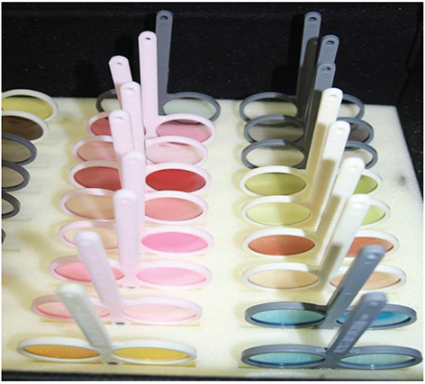
Fig. 1 MISViS filters consist of diverse colors of lenses. Each color family includes four to five lenses with differences in the degree of darkness.
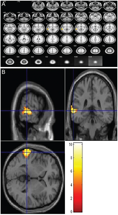
Fig. 2 Statistical parametric maps showing a significant blood-oxygenation-level-dependent activation in the left middle and superior temporal cortices in patients with Meares-Irlen syndrome during sentence reading with color-tinted lenses compared to reading without color-tinted lenses (paired t-test, p < 0.05, corrected for multiple comparisons using family wise error). The left side of each picture is the left side of the brain (A). The color bar represents the t-value (B). The images show mean differences in the activated regions before and after wearing different lenses.
Reference
-
1. Evans BJ, Wilkins AJ, Brown J, et al. A preliminary investigation into the aetiology of Meares-Irlen syndrome. Ophthalmic Physiol Opt. 1996; 16:286–296.2. Irlen H, Lass MJ. Improving reading problems due to symptoms of scotopic sensitivity syndrome using Irlen lenses and overlays. Education. 1989; 109:413–417.3. Evans BJ. The need for optometric investigation in suspected Meares-Irlen syndrome or visual stress. Ophthalmic Physiol Opt. 2005; 25:363–370.4. Loew SJ, Watson K. A prospective genetic marker of the visual-perception disorder Meares-Irlen syndrome. Percept Mot Skills. 2012; 114:870–882.5. Stein J. Visual motion sensitivity and reading. Neuropsychologia. 2003; 41:1785–1793.6. Kriss I, Evans BJ. The relationship between dyslexia and Meares-Irlen syndrome. J Res Read. 2005; 28:350–364.7. Kruk R, Sumbler K, Willows D. Visual processing characteristics of children with Meares-Irlen syndrome. Ophthalmic Physiol Opt. 2008; 28:35–46.8. Chang M, Kim SH, Kim JY, Cho YA. Specific visual symptoms and signs of Meares-Irlen syndrome in Korean. Korean J Ophthalmol. 2014; 28:159–163.9. Kim SH, Cho YA. Clinical characteristics of patients with dyslexia in Korea: correlation with meares-irlen syndrome. J Korean Ophthalmol Soc. 2010; 51:1639–1642.10. Park SH, Kim SH, Cho YA, Joo CK. The effect of colored filters in patients with Meares-Irlen syndrome. J Korean Ophthalmol Soc. 2012; 53:452–459.11. Logothetis NK. What we can do and what we cannot do with fMRI. Nature. 2008; 453:869–878.12. Friston KJ, Holmes AP, Poline JB, et al. Analysis of fMRI time-series revisited. Neuroimage. 1995; 2:45–53.13. Chase C, Ashourzadeh A, Kelly C, et al. Can the magnocellular pathway read? Evidence from studies of color. Vision Res. 2003; 43:1211–1222.14. O'Connor PD, Sofo F, Kendall L, Olsen G. Reading disabilities and the effects of colored filters. J Learn Disabil. 1990; 23:597–603.15. Robinson GL, Miles J. The use of coloured overlays to improve visual processing: a preliminary survey. Except Child. 1987; 34:65–70.16. Williams GJ, Kitchener G, Press LJ, et al. The use of tinted lenses and colored overlays for the treatment of dyslexia and other related reading and learning disorders. Optometry. 2004; 75:720–722.17. Friederici AD, Ruschemeyer SA, Hahne A, Fiebach CJ. The role of left inferior frontal and superior temporal cortex in sentence comprehension: localizing syntactic and semantic processes. Cereb Cortex. 2003; 13:170–177.18. Vandenberghe R, Nobre AC, Price CJ. The response of left temporal cortex to sentences. J Cogn Neurosci. 2002; 14:550–560.
- Full Text Links
-
- Actions
-
Cited
- CITED
-
- Close
- Share
-
- Similar articles
-
- Clinical Characteristics of Patients With Dyslexia in Korea : Correlation With Meares-Irlen Syndrome
- The Effect of Colored Filters in Patients with Meares-Irlen Syndrome
- Specific Visual Symptoms and Signs of Meares-Irlen Syndrome in Korean
- Functional Magnetic Resonance Imaging of the Brain: Principle and Practical Application
- Dynamic Contrast-Enhanced MR Imaging of Tietze’s Syndrome: a Case Report
