J Korean Med Sci.
2016 Feb;31(Suppl 1):S32-S37. 10.3346/jkms.2016.31.S1.S32.
Survey of Thoracic CT Protocols and Technical Parameters in Korean Hospitals: Changes before and after Establishment of Thoracic CT Guideline by Korean Society of Thoracic Radiology in 2008
- Affiliations
-
- 1Department of Radiology and Research Institute of Radiology, University of Ulsan College of Medicine, Seoul, Korea. dokh@amc.seoul.kr
- KMID: 2363378
- DOI: http://doi.org/10.3346/jkms.2016.31.S1.S32
Abstract
- We retrospectively reviewed the thoracic CT scan protocols and technical parameters obtained from hospitals in Korea, one group during May 2007 (n = 100) and the other group during January 2012 (n = 173), before and after the establishment of the thoracic CT Guideline in 2008. Each group was also divided into two subgroups according to the health care delivery level, i.e. the "A" subgroup from primary and the "B" subgroup from secondary and tertiary care hospitals. When comparing the data from 2007 and 2012, the tube current decreased from 179.1 mAs to 137.2 mAs. The scan interval decreased from 6.4 mm to 4.8 mm. Also, the insufficient scan range decreased from 19.0% to 8.7%, and the suboptimal quality scans decreased from 33.0% to 5.2%. Between groups A and B, group B had lower tube voltages, smaller scan thicknesses, and smaller scan intervals. However, group B had more phase numbers. In terms of the suboptimal quality scans, a decrease was seen in both groups. In conclusion, during the five-year time period between 2007 and 2012, a reduction in the tube current values was seen. And the overall image quality improved over the same time period. We assume that these changes are attributed to the implementation of the thoracic CT guideline in 2008.
Keyword
MeSH Terms
Figure
-
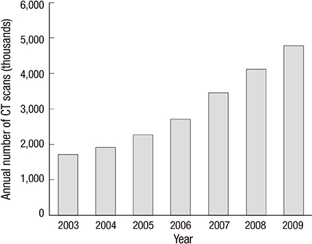
Fig. 1 The number of CT examinations in Korea between 2003 and 2009 (unit = 1,000).
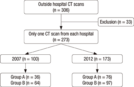
Fig. 2 The flow of this study.
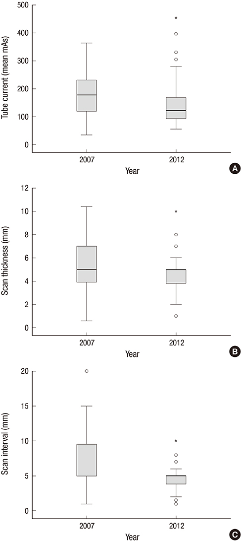
Fig. 3 A comparison between 2007 and 2012. (A) Tube current. (B) Scan thickness. (C) Scan interval. °, outliers; *, extreme values.
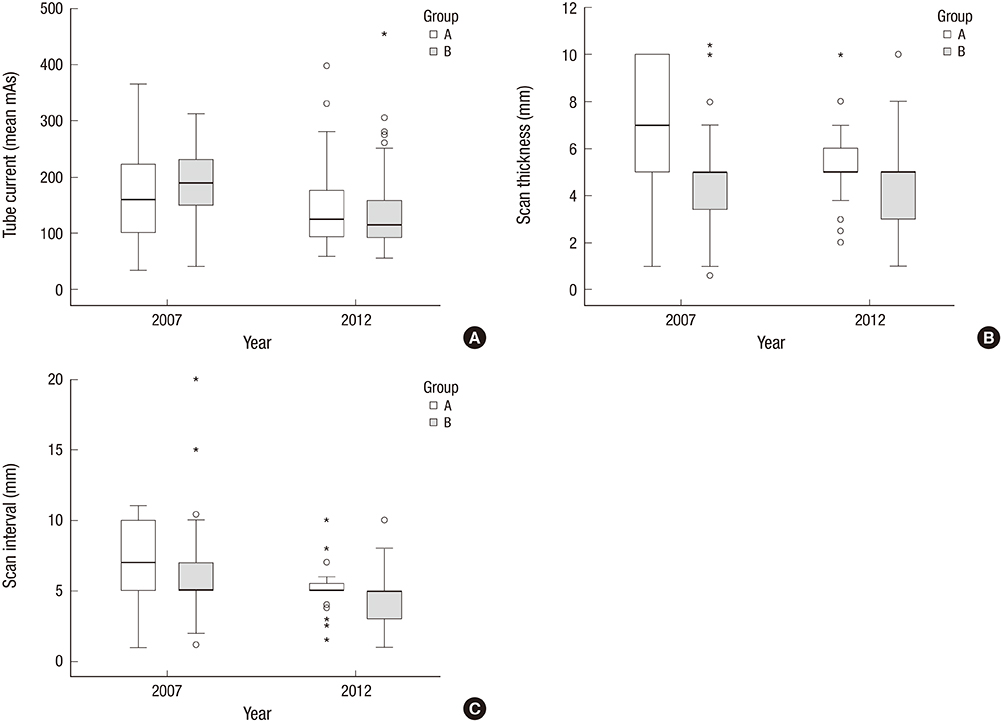
Fig. 4 A comparison between group A and B. (A) Tube current. (B) Scan thickness. (C) Scan interval. °, outliers; *, extreme values.
Reference
-
1. Mettler FA Jr, Bhargavan M, Faulkner K, Gilley DB, Gray JE, Ibbott GS, Lipoti JA, Mahesh M, McCrohan JL, Stabin MG, et al. Radiologic and nuclear medicine studies in the United States and worldwide: frequency, radiation dose, and comparison with other radiation sources--1950-2007. Radiology. 2009; 253:520–531.2. American College of Radiology. ACR practice guideline for the performance of pediatric and adult thoracic computed tomography(CT). accessed on 15 October 2015. Available at http://www.acr.org.3. Brenner DJ, Doll R, Goodhead DT, Hall EJ, Land CE, Little JB, Lubin JH, Preston DL, Preston RJ, Puskin JS, et al. Cancer risks attributable to low doses of ionizing radiation: assessing what we really know. Proc Natl Acad Sci U S A. 2003; 100:13761–13766.4. Multislice Computed Tomography. 2004 CT quality criteria. accessed on 15 October 2015. Available at http://msct.eu/CT_Quality_Criteria.htm.5. Ministry for Health, Welfare and Family Affairs. National Institute of Food and Drug Safety Evaluation. The Korean Radiological Society. The Korean Radiological Technologists Association. Guideline for diagnostic reference level of the radiation exposure of CT examination. accessed on 15 October 2015. Available at http://www.mfds.go.kr/medicaldevice/index.do?nMenuCode=121&page=1&categoryCode1=86&page=1&mode=view&boardSeq=49621.6. Health Insurance Review and Assessment Service. Assessments of the 2006 state of CT claims reported by the Korea Health Insurance Review and Assessment Service. accessed on 15 October 2015. Available at http://biz.hira.or.kr/ICSFiles/afieldfile/2008/01/21/2006_CT.pdf.7. Brenner DJ, Hall EJ. Computed tomography--an increasing source of radiation exposure. N Engl J Med. 2007; 357:2277–2284.8. Hricak H, Brenner DJ, Adelstein SJ, Frush DP, Hall EJ, Howell RW, McCollough CH, Mettler FA Jr, Pearce MS, Suleiman OH, et al. Managing radiation use in medical imaging: a multifaceted challenge. Radiology. 2011; 258:889–905.9. National Council on Radiation Protection and Measurements. Ionizing radiation exposure of the population of the United States: NCRP Report No.160. Bethesda, MD: National Council on Radiation Protection and Measurements;2009.10. Hall EJ, Brenner DJ. Cancer risks from diagnostic radiology. Br J Radiol. 2008; 81:362–378.11. Tsushima Y, Taketomi-Takahashi A, Takei H, Otake H, Endo K. Radiation exposure from CT examinations in Japan. BMC Med Imaging. 2010; 10:24.12. Kim BH, Do KH, Goo HW, Yang DH, Oh SY, Kim HJ, Lee KY, Lee JE. National survey of radiation doses of pediatric chest radiography in Korea: analysis of the factors affecting radiation doses. Korean J Radiol. 2012; 13:610–617.13. Tanimura M, Matsui I, Abe J, Ikeda H, Kobayashi N, Ohira M, Yokoyama M, Kaneko M. Increased risk of hepatoblastoma among immature children with a lower birth weight. Cancer Res. 1998; 58:3032–3035.14. Shu XO, Potter JD, Linet MS, Severson RK, Han D, Kersey JH, Neglia JP, Trigg ME, Robison LL. Diagnostic X-rays and ultrasound exposure and risk of childhood acute lymphoblastic leukemia by immunophenotype. Cancer Epidemiol Biomarkers Prev. 2002; 11:177–185.15. Shu XO, Jin F, Linet MS, Zheng W, Clemens J, Mills J, Gao YT. Diagnostic X-ray and ultrasound exposure and risk of childhood cancer. Br J Cancer. 1994; 70:531–536.16. Health and Safety at Work etc. Act 1974. accessed on 15 October 2015. Available at http://www.legislation.gov.uk/ukpga/1974/37/pdfs/ukpga_19740037_en.pdf.17. Goo HW. CT radiation dose optimization and estimation: an update for radiologists. Korean J Radiol. 2012; 13:1–11.18. Kalra MK, Maher MM, Toth TL, Hamberg LM, Blake MA, Shepard JA, Saini S. Strategies for CT radiation dose optimization. Radiology. 2004; 230:619–628.19. Singh S, Kalra MK, Moore MA, Shailam R, Liu B, Toth TL, Grant E, Westra SJ. Dose reduction and compliance with pediatric CT protocols adapted to patient size, clinical indication, and number of prior studies. Radiology. 2009; 252:200–208.20. Korean Society of Thoracic Radiology Guidelines Standards Committee. KSTR chest CT practice guidelines and technical standards. accessed on 15 October 2015. Available at http://kstr.radiology.or.kr/data/policy.html.21. Shrimpton PC, Hillier MC, Meeson S, Golding SJ. Doses from computed tomography(CT) examinations in the UK: 2011 review. London: Public Health England;2014.22. Brix G, Nagel HD, Stamm G, Veit R, Lechel U, Griebel J, Galanski M. Radiation exposure in multi-slice versus single-slice spiral CT: results of a nationwide survey. Eur Radiol. 2003; 13:1979–1991.23. van der Molen AJ, Schilham A, Stoop P, Prokop M, Geleijns J. A national survey on radiation dose in CT in the Netherlands. Insights Imaging. 2013; 4:383–390.24. Tsai HY, Tung CJ, Yu CC, Tyan YS. Survey of computed tomography scanners in Taiwan: dose descriptors, dose guidance levels, and effective doses. Med Phys. 2007; 34:1234–1243.25. Suliman II, Abdalla SE, Ahmed NA, Galal MA, Salih I. Survey of computed tomography technique and radiation dose in Sudanese hospitals. Eur J Radiol. 2011; 80:e544–51.26. Wambani JS, Korir GK, Onditi EG, Korir IK. A survey of computed tomography imaging techniques and patient dose in Kenya. East Afr Med J. 2010; 87:400–407.27. Ogbole GI, Obed R. Radiation doses in computed tomography: need for optimization and application of dose reference levels in Nigeria. West Afr J Radiol. 2014; 21:1–6.
