J Korean Med Sci.
2016 Feb;31(Suppl 1):S24-S31. 10.3346/jkms.2016.31.S1.S24.
Radiation Doses of Various CT Protocols: a Multicenter Longitudinal Observation Study
- Affiliations
-
- 1Department of Radiology, Seoul St. Mary's Hospital, College of Medicine, the Catholic University of Korea, Seoul, Korea. sejung@catholic.ac.kr
- 2Department of Radiology, Samsung Medical Center, Sungkyunkwan University, School of Medicine, Seoul, Korea.
- 3Department of Radiology, Bucheon St. Mary's Hospital, College of Medicine, the Catholic University of Korea, Bucheon, Korea.
- 4Department of Radiology, Hanyang University Guri Hospital, College of Medicine, Hanyang University, Guri, Korea.
- 5Department of Radiology, Severance Hospital, College of Medicine, Yonsei University, Seoul, Korea.
- 6Department of Radiology, Gangnam Severance Hospital, College of Medicine, Yonsei University, Seoul, Korea.
- 7Department of Radiology, Korea University Guro Hospital, College of Medicine, Korea University, Seoul, Korea.
- 8Department of Radiology, St. Vincent's Hospital, College of Medicine, the Catholic University of Korea, Suwon, Korea.
- KMID: 2363377
- DOI: http://doi.org/10.3346/jkms.2016.31.S1.S24
Abstract
- Emerging concerns regarding the hazard from medical radiation including CT examinations has been suggested. The purpose of this study was to observe the longitudinal changes of CT radiation doses of various CT protocols and to estimate the long-term efforts of supervising radiologists to reduce medical radiation. Radiation dose data from 11 representative CT protocols were collected from 12 hospitals. Attending radiologists had collected CT radiation dose data in two time points, 2007 and 2010. They collected the volume CT dose index (CTDIvol) of each phase, number of phases, dose length product (DLP) of each phase, and types of scanned CT machines. From the collected data, total DLP and effective dose (ED) were calculated. CTDIvol, total DLP, and ED of 2007 and 2010 were compared according to CT protocols, CT machine type, and hospital. During the three years, CTDIvol had significantly decreased, except for dynamic CT of the liver. Total DLP and ED were significantly decreased in all 11 protocols. The decrement was more evident in newer CT scanners. However, there was substantial variability of changes of ED during the three years according to hospitals. Although there was variability according to protocols, machines, and hospital, CT radiation doses were decreased during the 3 years. This study showed the effects of decreased CT radiation dose by efforts of radiologists and medical society.
MeSH Terms
Figure
-
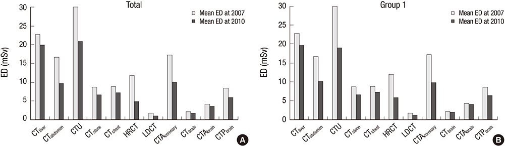
Fig. 1 Mean effective doses according to 11 CT protocols. (A) Total data comparison between 2007 and 2010. (B) Comparison from the data of same CT scanners from 2007 and 2010 (group 1).

Fig. 2 Percentage changes of the volume CT dose index (CTDIvol) and total dose-length product (DLP) of 11 CT protocols. (A) Total data comparison between 2007 and 2010. (B) Comparison from data of same CT scanners from 2007 and 2010 (group 1). Note the discrepancies between reduction of CTDIvol and total DLP in CTliver, CTabdomen, and HRCT. A similar trend was observed in group 1.
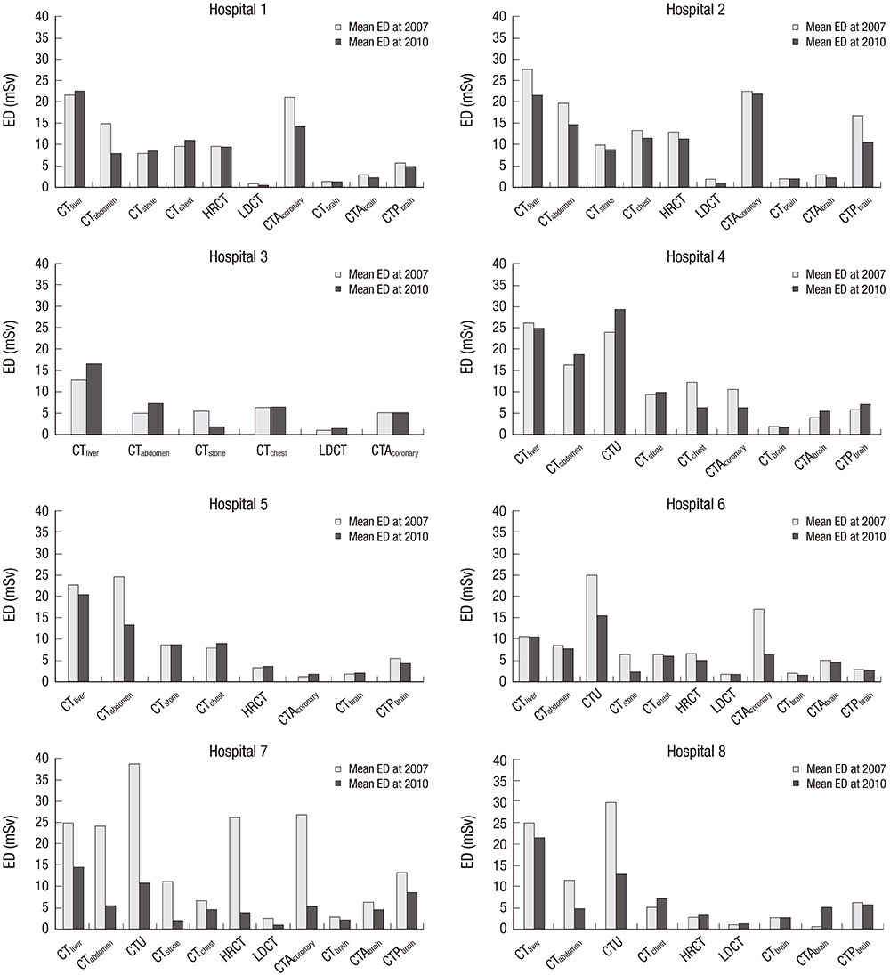
Fig. 3 Changes of mean effective doses of 8 hospitals in group 1. Note the variability of EDs at 2007 and changes during 3 years according to the protocols.
Reference
-
1. Brenner DJ, Hall EJ. Computed tomography--an increasing source of radiation exposure. N Engl J Med. 2007; 357:2277–2284.2. Berrington de González A, Mahesh M, Kim KP, Bhargavan M, Lewis R, Mettler F, Land C. Projected cancer risks from computed tomographic scans performed in the United States in 2007. Arch Intern Med. 2009; 169:2071–2077.3. Kalra MK, Maher MM, Toth TL, Hamberg LM, Blake MA, Shepard JA, Saini S. Strategies for CT radiation dose optimization. Radiology. 2004; 230:619–628.4. Shrimpton PC, Hillier MC, Lewis MA, Dunn M. National survey of doses from CT in the UK: 2003. Br J Radiol. 2006; 79:968–980.5. Tsapaki V, Aldrich JE, Sharma R, Staniszewska MA, Krisanachinda A, Rehani M, Hufton A, Triantopoulou C, Maniatis PN, Papailiou J, et al. Dose reduction in CT while maintaining diagnostic confidence: diagnostic reference levels at routine head, chest, and abdominal CT--IAEA-coordinated research project. Radiology. 2006; 240:828–834.6. Huda W, Mettler FA. Volume CT dose index and dose-length product displayed during CT: what good are they? Radiology. 2011; 258:236–242.7. Yoon MA, Kim SH, Lee JM, Woo HS, Lee ES, Ahn SJ, Han JK. Adaptive statistical iterative reconstruction and Veo: assessment of image quality and diagnostic performance in CT colonography at various radiation doses. J Comput Assist Tomogr. 2012; 36:596–601.8. Schuhbaeck A, Achenbach S, Layritz C, Eisentopf J, Hecker F, Pflederer T, Gauss S, Rixe J, Kalender W, Daniel WG, et al. Image quality of ultra-low radiation exposure coronary CT angiography with an effective dose <0.1 mSv using high-pitch spiral acquisition and raw data-based iterative reconstruction. Eur Radiol. 2013; 23:597–606.9. Yamada Y, Jinzaki M, Hosokawa T, Tanami Y, Sugiura H, Abe T, Kuribayashi S. Dose reduction in chest CT: comparison of the adaptive iterative dose reduction 3D, adaptive iterative dose reduction, and filtered back projection reconstruction techniques. Eur J Radiol. 2012; 81:4185–4195.10. De Cecco CN, Darnell A, Macías N, Ayuso JR, Rodríguez S, Rimola J, Pagés M, García-Criado A, Rengo M, Laghi A, et al. Virtual unenhanced images of the abdomen with second-generation dual-source dual-energy computed tomography: image quality and liver lesion detection. Invest Radiol. 2013; 48:1–9.11. Jessen KA, Shrimpton PC, Geleijns J, Panzer W, Tosi G. Dosimetry for optimisation of patient protection in computed tomography. Appl Radiat Isot. 1999; 50:165–172.12. Bongartz G, Golding SJ, Jurik AG, Leonardi M, van Persijn van Meerten E, Rodríguez R, Schneider K, Calzado A, Geleijns J, Jessen KA, et al. European guidelines for multislice computed tomography: appendix A. Brussels: European Guidelines for Multislice Computed Tomography Funded by the European Commission;2004.13. Hara AK, Paden RG, Silva AC, Kujak JL, Lawder HJ, Pavlicek W. Iterative reconstruction technique for reducing body radiation dose at CT: feasibility study. AJR Am J Roentgenol. 2009; 193:764–771.14. Sung DW. A study of strategies for patient dose management. Seoul: Ministry of Health and Welfare;2012.15. Van Der Molen AJ, Cowan NC, Mueller-Lisse UG, Nolte-Ernsting CC, Takahashi S, Cohan RH. CT Urography Working Group of the European Society of Urogenital Radiology (ESUR). CT urography: definition, indications and techniques. A guideline for clinical practice. Eur Radiol. 2008; 18:4–17.16. Lee CH, Goo JM, Ye HJ, Ye SJ, Park CM, Chun EJ, Im JG. Radiation dose modulation techniques in the multidetector CT era: from basics to practice. Radiographics. 2008; 28:1451–1459.17. Smith AB, Dillon WP, Gould R, Wintermark M. Radiation dose-reduction strategies for neuroradiology CT protocols. AJNR Am J Neuroradiol. 2007; 28:1628–1632.18. Linton OW, Mettler FA Jr; National Council on Radiation Protection and Measurements. National conference on dose reduction in CT, with an emphasis on pediatric patients. AJR Am J Roentgenol. 2003; 181:321–329.19. Tamm EP, Rong XJ, Cody DD, Ernst RD, Fitzgerald NE, Kundra V. Quality initiatives: CT radiation dose reduction: how to implement change without sacrificing diagnostic quality. Radiographics. 2011; 31:1823–1832.20. McCollough CH, Bruesewitz MR, Kofler JM Jr. CT dose reduction and dose management tools: overview of available options. Radiographics. 2006; 26:503–512.21. McCollough CH, Primak AN, Braun N, Kofler J, Yu L, Christner J. Strategies for reducing radiation dose in CT. Radiol Clin North Am. 2009; 47:27–40.22. Antypas EJ, Sokhandon F, Farah M, Emerson S, Bis KG, Tien H, Mezwa D. A comprehensive approach to CT radiation dose reduction: one institution’s experience. AJR Am J Roentgenol. 2011; 197:935–940.23. Wallace AB, Goergen SK, Schick D, Soblusky T, Jolley D. Multidetector CT dose: clinical practice improvement strategies from a successful optimization program. J Am Coll Radiol. 2010; 7:614–624.
- Full Text Links
-
- Actions
-
Cited
- CITED
-
- Close
- Share
-
- Similar articles
-
- CT radiation dose and radiation reduction strategies
- CT Examinations for COVID-19: A Systematic Review of Protocols, Radiation Dose, and Numbers Needed to Diagnose and Predict
- Comparison of Chest Pain Protocols for Electrocardiography-Gated Dual-Source Cardiothoracic CT in Children and Adults: The Effect of Tube Current Saturation on Radiation Dose Reduction
- Strategies of computed tomography radiation dose reduction: justification and optimization
- Comparison of Image Quality of Shoulder CT Arthrography Conducted Using 120 kVp and 140 kVp Protocols
