Interposition of the Posterior Cruciate Ligament into the Medial Compartment of the Knee Joint on Coronal Magnetic Resonance Imaging
- Affiliations
-
- 1Department of Radiology, Samsung Medical Center, Sungkyunkwan University School of Medicine, Seoul 06351, Korea. youngcheol.yoon@gmail.com
- 2Department of Medical Device Management and Research, SAIHST, Sungkyunkwan University, Seoul 06351, Korea.
- 3Department of Orthopedic Surgery, Samsung Medical Center, Sungkyunkwan University School of Medicine, Seoul 06351, Korea.
- 4Department of Preventive Medicine, Kyung Hee University School of Medicine, Seoul 02454, Korea.
- KMID: 2360209
- DOI: http://doi.org/10.3348/kjr.2016.17.2.239
Abstract
OBJECTIVE
The purpose of our study was to evaluate the overall prevalence and clinical significance of interposition of the posterior cruciate ligament (PCL) into the medial compartment of the knee joint in coronal magnetic resonance imaging (MRI).
MATERIALS AND METHODS
We retrospectively reviewed 317 consecutive patients referred for knee MRI at our institution between October 2009 and December 2009. Interposition of the PCL into the medial compartment of the knee joint on proton coronal MRI was evaluated dichotomously (i.e., present or absent). We analyzed the interposition according to its prevalence as well as its relationship with right-left sidedness, gender, age, and disease categories (osteoarthritis, anterior cruciate ligament tear, and medial meniscus tear).
RESULTS
Prevalence of interposition of PCL into the medial compartment of the knee joint was 47.0% (149/317). There was no right (50.0%, 83/166) to left (43.7%, 66/151) or male (50.3%, 87/173) to female (43.1%, 62/144) differences in the prevalence. There was no significant association between the prevalence and age, or the disease categories.
CONCLUSION
Interposition of the PCL into the medial compartment of the knee joint is observed in almost half of patients on proton coronal MRI of the knee. Its presence is not associated with any particular factors including knee pathology and may be regarded as a normal MR finding.
MeSH Terms
Figure
-
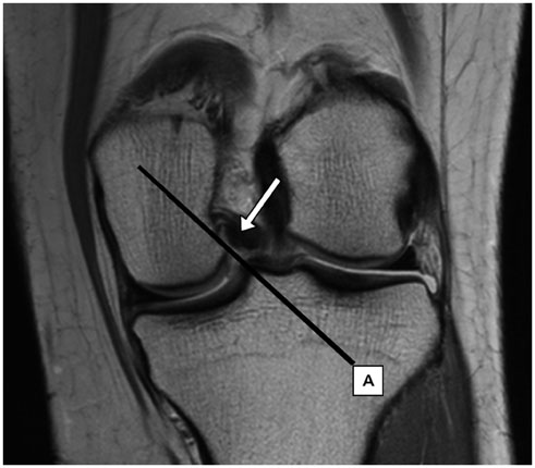
Fig. 1 47-year-old woman with low grade chondral lesion at retro-patellar cartilage. Proton density weighted (repetition time, 2200 msec; echo time, 30 msec) coronal MR image of left knee. Medial margin of PCL is located lateral to line between inferomedial corner of medial femoral condyle and medial tibial spine (arrow). This case was classified as negative for interposition of PCL into medial compartment of knee joint. PCL = posterior cruciate ligament
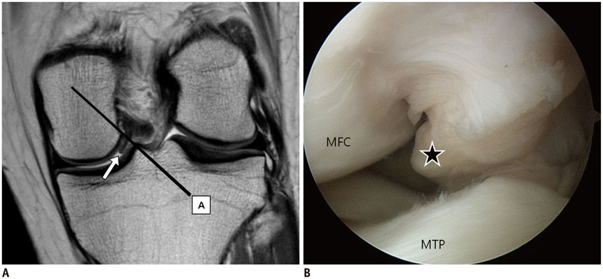
Fig. 2 44-year-old woman with partial tear of anterior cruciate ligament. Proton density weighted (repetition time, 2200 msec; echo time, 30 msec) coronal MR image (A) of left knee shows focal outward bulging contour at medial margin of PCL, confined to medial side of line between inferomedial corner of medial femoral condyle and medial tibial spine (arrow). This case was classified as positive for interposition of PCL into medial compartment of knee joint. Arthroscopic image of medial compartment obtained with knee flexion position (B) shows same focal outward bulging contour of PCL (asterisk) between medial femoral condyle (MFC) and medial tibial plateau (MTP). PCL = posterior cruciate ligament
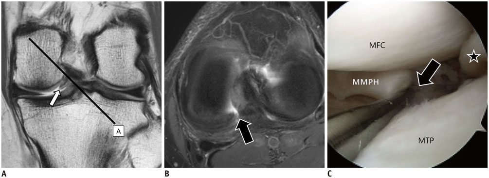
Fig. 3 49-year-old man with transverse tear of medial meniscus. Proton density weighted (repetition time, 2200 msec; echo time, 30 msec) coronal MR image (A) of left knee shows focal outward bulging contour at medial margin of PCL, confined to medial side of line between inferomedial corner of medial femoral condyle and medial tibial spine (arrow). This case was classified as positive for interposition of PCL into medial compartment of knee joint. Fat suppressed proton density weighted (repetition time, 2530 msec; echo time, 30 msec) axial MR image (B) shows full thickness transverse tear of medial meniscus posterior horn. Arthroscopic image of medial compartment obtained with knee flexion position (C) shows focal outward bulging contour of PCL (asterisk) between medial femoral condyle (MFC) and medial tibial plateau (MTP) and transverse tear of medial meniscus posterior horn (MMPH) (arrow). PCL = posterior cruciate ligament
Cited by 1 articles
-
Clinical Feasibility of Synthetic Magnetic Resonance Imaging in the Diagnosis of Internal Derangements of the Knee
Jisook Yi, Young Han Lee, Ho-Taek Song, Jin-Suck Suh
Korean J Radiol. 2018;19(2):311-319. doi: 10.3348/kjr.2018.19.2.311.
Reference
-
1. Sonin AH, Fitzgerald SW, Hoff FL, Friedman H, Bresler ME. MR imaging of the posterior cruciate ligament: normal, abnormal, and associated injury patterns. Radiographics. 1995; 15:551–561.2. Kim HK, Laor T, Shire NJ, Bean JA, Dardzinski BJ. Anterior and posterior cruciate ligaments at different patient ages: MR imaging findings. Radiology. 2008; 247:826–835.3. Turner DA, Prodromos CC, Petasnick JP, Clark JW. Acute injury of the ligaments of the knee: magnetic resonance evaluation. Radiology. 1985; 154:717–722.4. Rodriguez W Jr, Vinson EN, Helms CA, Toth AP. MRI appearance of posterior cruciate ligament tears. AJR Am J Roentgenol. 2008; 191:1031.5. Fitzgerald SW, Remer EM, Friedman H, Rogers LF, Hendrix RW, Schafer MF. MR evaluation of the anterior cruciate ligament: value of supplementing sagittal images with coronal and axial images. AJR Am J Roentgenol. 1993; 160:1233–1237.6. Roberts CC, Towers JD, Spangehl MJ, Carrino JA, Morrison WB. Advanced MR imaging of the cruciate ligaments. Radiol Clin North Am. 2007; 45:1003–1016. vi–vii.7. Stoller DW. Magnetic resonance imaging in orthopaedics and sports medicine. 3rd ed. Baltimore: Lippincott Williams & Wilkins;2007.8. Chen WT, Shih TT, Tu HY, Chen RC, Shau WY. Partial and complete tear of the anterior cruciate ligament. Acta Radiol. 2002; 43:511–516.9. Bolog N, Hodler J. MR imaging of the posterolateral corner of the knee. Skeletal Radiol. 2007; 36:715–728.10. Ogden JA. Injury to the growth mechanisms of the immature skeleton. Skeletal Radiol. 1981; 6:237–253.11. Robertson PL, Schweitzer ME, Bartolozzi AR, Ugoni A. Anterior cruciate ligament tears: evaluation of multiple signs with MR imaging. Radiology. 1994; 193:829–834.12. Boeree NR, Ackroyd CE. Magnetic resonance imaging of anterior cruciate ligament rupture. A new diagnostic sign. J Bone Joint Surg Br. 1992; 74:614–616.13. Akisue T, Stulberg BN, Bauer TW, McMahon JT, Wilde AH, Kurosaka M. Histologic evaluation of posterior cruciate ligaments from osteoarthritic knees. Clin Orthop Relat Res. 2002; (400):165–173.14. Mullaji AB, Marawar SV, Simha M, Jindal G. Cruciate ligaments in arthritic knees: a histologic study with radiologic correlation. J Arthroplasty. 2008; 23:567–572.15. Roemer FW, Kassim Javaid M, Guermazi A, Thomas M, Kiran A, Keen R, et al. Anatomical distribution of synovitis in knee osteoarthritis and its association with joint effusion assessed on non-enhanced and contrast-enhanced MRI. Osteoarthritis Cartilage. 2010; 18:1269–1274.16. Fanelli GC, Beck JD, Edson CJ. Current concepts review: the posterior cruciate ligament. J Knee Surg. 2010; 23:61–72.17. Fox RJ, Harner CD, Sakane M, Carlin GJ, Woo SL. Determination of the in situ forces in the human posterior cruciate ligament using robotic technology. A cadaveric study. Am J Sports Med. 1998; 26:395–340.18. Ortiz GJ, Schmotzer H, Bernbeck J, Graham S, Tibone JE, Vangsness CT Jr. Isometry of the posterior cruciate ligament. Effects of functional load and muscle force application. Am J Sports Med. 1998; 26:663–668.19. Osti M, Tschann P, Künzel KH, Benedetto KP. Anatomic characteristics and radiographic references of the anterolateral and posteromedial bundles of the posterior cruciate ligament. Am J Sports Med. 2012; 40:1558–1563.20. Edwards A, Bull AM, Amis AA. The attachments of the fiber bundles of the posterior cruciate ligament: an anatomic study. Arthroscopy. 2007; 23:284–290.21. Gali JC, Esquerdo P, Almagro MA, da Silva PA. Radiographic study on the tibial insertion of the posterior cruciate ligament. Rev Bras Ortop. 2015; 50:342–347.
- Full Text Links
-
- Actions
-
Cited
- CITED
-
- Close
- Share
-
- Similar articles
-
- A Clinical Study of Acute Posterior Cruciate Ligament Injury
- Reconstruction of the Posterior Cruciate Ligament Using the Medial Meniscus
- Ganglion Cyst of the Posterior Cruciate Ligament: A Case Report
- Intraarticular Ganglion Arising from the Posterior Cruciate Ligament
- Reconstruction of Posterior Cruciate Ligament Using Semitendinosus Tendon (3 cases)
