Detecting the Progression of Normal Tension Glaucoma: A Comparison of Perimetry, Optic Coherence Tomography, and Heidelberg Retinal Tomography
- Affiliations
-
- 1Department of Ophthalmology, Saevit Eye Hospital, Goyang, Korea.
- 2Department of Ophthalmology and Visual Science, Seoul St. Mary's Hospital, The Catholic University of Korea College of Medicine, Seoul, Korea. ckpark@catholic.ac.kr
- KMID: 2360135
- DOI: http://doi.org/10.3341/kjo.2015.29.1.31
Abstract
- PURPOSE
We compared the abilities of Stratus optical coherence tomography (OCT), Heidelberg retinal tomography (HRT) and standard automated perimetry (SAP) to detect the progression of normal tension glaucoma (NTG) in patients whose eyes displayed localized retinal nerve fiber layer (RNFL) defect enlargements.
METHODS
One hundred four NTG patients were selected who met the selection criteria: a localized RNFL defect visible on red-free fundus photography, a minimum of five years of follow-up, and a minimum of five reliable SAP, Stratus OCT and HRT tests. Tests which detected progression at any visit during the 5-year follow-up were identified, and patients were further classified according to the state of the glaucoma using the mean deviation (MD) of SAP. For each test, the overall rates of change were calculated for parameters that differed significantly between patients with and without NTG progression.
RESULTS
Forty-seven (45%) out of 104 eyes displayed progression that could be detected by red-free fundus photography. Progression was detected in 27 (57%) eyes using SAP, 19 (40%) eyes using OCT, and 17 (36%) eyes using HRT. In early NTG, SAP detected progression in 44% of eyes, and this increased to 70% in advanced NTG. In contrast, OCT and HRT detected progression in 50 and 7% of eyes during early NTG, but only 30 and 0% of eyes in advanced NTG, respectively. Among several parameters, the rates of change that differed significantly between patients with and without progression were the MD of SAP (p = 0.013), and the inferior RNFL thickness (p = 0.041) and average RNFL thickness (p = 0.032) determined by OCT.
CONCLUSIONS
SAP had a higher detection rate of NTG progression than other tests, especially in patients with advanced glaucoma, when we defined progression as the enlargement of a localized RNFL defect. The rates of change of the MD of SAP, inferior RNFL thickness, and average RNFL thickness differed between NTG patients with and without progression.
Keyword
MeSH Terms
Figure
-

Fig. 1 Detection rate of progression by each diagnostic test in 47 patients having an eye with normal tension glaucoma that displayed progression, defined as enlargement of a localized retinal nerve fiber layer defect on red-free fundus photography. SAP = standard automated perimetry; HRT = Heidelberg retinal tomography; OCT = optical coherence tomography.
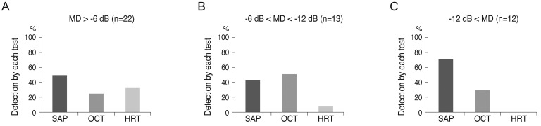
Fig. 2 Comparison of methods for detecting disease progression. Sample included 47 patients with normal tension glaucoma who were assessed for enlargement of the localized retinal nerve fiber layer by red-free fundus photography. Data shows detecting disease progression according to glaucoma severity (A, mild glaucoma; B, moderate glaucoma; C, severe glaucoma). MD = mean deviation; SAP = standard automated perimetry; OCT = optical coherence tomography; HRT = Heidelberg retinal tomography.
Reference
-
1. Anderson DR, Drance SM, Schulzer M. Collaborative Normal-Tension Glaucoma Study Group. Natural history of normal-tension glaucoma. Ophthalmology. 2001; 108:247–253. PMID: 11158794.2. Johnson CA, Sample PA, Zangwill LM, et al. Structure and function evaluation (SAFE). II. Comparison of optic disk and visual field characteristics. Am J Ophthalmol. 2003; 135:148–154. PMID: 12566017.
Article3. Quigley HA, Katz J, Derick RJ, et al. An evaluation of optic disc and nerve fiber layer examinations in monitoring progression of early glaucoma damage. Ophthalmology. 1992; 99:19–28. PMID: 1741133.
Article4. Sommer A, Katz J, Quigley HA, et al. Clinically detectable nerve fiber atrophy precedes the onset of glaucomatous field loss. Arch Ophthalmol. 1991; 109:77–83. PMID: 1987954.
Article5. Nouri-Mahdavi K, Hoffman D, Ralli M, Caprioli J. Comparison of methods to predict visual field progression in glaucoma. Arch Ophthalmol. 2007; 125:1176–1181. PMID: 17846355.
Article6. Chang RT, Budenz DL. Diagnosing glaucoma progression. Int Ophthalmol Clin. 2008; 48:13–28. PMID: 18936634.
Article7. Kass MA, Heuer DK, Higginbotham EJ, et al. The Ocular Hypertension Treatment Study: a randomized trial determines that topical ocular hypotensive medication delays or prevents the onset of primary open-angle glaucoma. Arch Ophthalmol. 2002; 120:701–713. PMID: 12049574.8. Medeiros FA, Zangwill LM, Bowd C, et al. Use of progressive glaucomatous optic disk change as the reference standard for evaluation of diagnostic tests in glaucoma. Am J Ophthalmol. 2005; 139:1010–1018. PMID: 15953430.
Article9. Miglior S, Zeyen T, Pfeiffer N, et al. Results of the European glaucoma prevention study. Ophthalmology. 2005; 112:366–375. PMID: 15745761.10. Weinreb RN, Lusky M, Bartsch DU, Morsman D. Effect of repetitive imaging on topographic measurements of the optic nerve head. Arch Ophthalmol. 1993; 111:636–638. PMID: 8489444.
Article11. Gloor B, Schmied U, Faessler A. Changes of glaucomatous field defects: degree of accuracy of measurements with the automatic perimeter Octopus. Int Ophthalmol. 1980; 3:5–10. PMID: 7012057.12. Leske MC, Heijl A, Hussein M, et al. Factors for glaucoma progression and the effect of treatment: the early manifest glaucoma trial. Arch Ophthalmol. 2003; 121:48–56. PMID: 12523884.13. Quigley HA, Reacher M, Katz J, et al. Quantitative grading of nerve fiber layer photographs. Ophthalmology. 1993; 100:1800–1807. PMID: 8259277.
Article14. Suh MH, Kim DM, Kim YK, et al. Patterns of progression of localized retinal nerve fibre layer defect on red-free fundus photographs in normal-tension glaucoma. Eye (Lond). 2010; 24:857–863. PMID: 19680281.
Article15. Schulzer M. Errors in the diagnosis of visual field progression in normal-tension glaucoma. Ophthalmology. 1994; 101:1589–1594. PMID: 8090461.16. Wollstein G, Schuman JS, Price LL, et al. Optical coherence tomography longitudinal evaluation of retinal nerve fiber layer thickness in glaucoma. Arch Ophthalmol. 2005; 123:464–470. PMID: 15824218.
Article17. Arthur SN, Smith SD, Wright MM, et al. Reproducibility and agreement in evaluating retinal nerve fibre layer thickness between Stratus and Spectralis OCT. Eye (Lond). 2011; 25:192–200. PMID: 21109776.
Article18. Toteberg-Harms M, Sturm V, Knecht PB, et al. Repeatability of nerve fiber layer thickness measurements in patients with glaucoma and without glaucoma using spectral-domain and time-domain OCT. Graefes Arch Clin Exp Ophthalmol. 2012; 250:279–287. PMID: 21909812.
Article19. Vizzeri G, Weinreb RN, Martinez de la Casa JM, et al. Clinicians agreement in establishing glaucomatous progression using the Heidelberg retina tomograph. Ophthalmology. 2009; 116:14–24. PMID: 19010552.
Article20. Jonas JB, Grundler AE. Correlation between mean visual field loss and morphometric optic disk variables in the open-angle glaucomas. Am J Ophthalmol. 1997; 124:488–497. PMID: 9323939.
Article21. Bartz-Schmidt KU, Thumann G, Jonescu-Cuypers CP, Krieglstein GK. Quantitative morphologic and functional evaluation of the optic nerve head in chronic open-angle glaucoma. Surv Ophthalmol. 1999; 44 Suppl 1:S41–S53. PMID: 10548116.
Article22. Sample PA, Johnson CA. Functional assessment of glaucoma. J Glaucoma. 2001; 10:S49–S52. PMID: 11890275.
Article23. Girkin CA. Relationship between structure of optic nerve/nerve fiber layer and functional measurements in glaucoma. Curr Opin Ophthalmol. 2004; 15:96–101. PMID: 15021219.
Article24. Harwerth RS, Carter-Dawson L, Shen F, et al. Ganglion cell losses underlying visual field defects from experimental glaucoma. Invest Ophthalmol Vis Sci. 1999; 40:2242–2250. PMID: 10476789.25. Garway-Heath DF, Holder GE, Fitzke FW, Hitchings RA. Relationship between electrophysiological, psychophysical, and anatomical measurements in glaucoma. Invest Ophthalmol Vis Sci. 2002; 43:2213–2220. PMID: 12091419.
- Full Text Links
-
- Actions
-
Cited
- CITED
-
- Close
- Share
-
- Similar articles
-
- Correlation between Heidelberg Retina Tomograph, Visual Field, and Optical Coherence Tomography in Primary Open-Angle Glaucoma
- Comparison of OCT and HRT Findings Among Normal, Normal Tension Glaucoma, and High Tension Glaucoma
- Comparison of Cup-to-disc Ratio Using the Superfield Lens and Optical Coherence Tomography
- Correlation Between Disc Size and Retinal Nerve Fiber Layer Thickness in Normal Tension Glaucoma
- Correlation between Retinal Nerve Fiber Layer Thickness and Visual Field in Normal Tension Glaucoma Patients
