Transient Corneal Edema is a Predictive Factor for Pseudophakic Cystoid Macular Edema after Uncomplicated Cataract Surgery
- Affiliations
-
- 1Department of Ophthalmology, Dongguk University Ilsan Hospital, Goyang, Korea. oph0112@gmail.com
- 2Department of Ophthalmology and Visual Sciences, Montefiore Medical Center, Albert Einstein College of Medicine, Bronx, NY, USA.
- KMID: 2360133
- DOI: http://doi.org/10.3341/kjo.2015.29.1.14
Abstract
- PURPOSE
To report transient corneal edema after phacoemulsification as a predictive factor for the development of pseudophakic cystoid macular edema (PCME).
METHODS
A total of 150 eyes from 150 patients (59 men and 91 women; mean age, 68.0 ± 10.15 years) were analyzed using spectral domain optical coherence tomography 1 week and 5 weeks after routine phacoemulsification cataract surgery. Transient corneal edema detected 1 week after surgery was analyzed to reveal any significant relationship with the development of PCME 5 weeks after surgery.
RESULTS
Transient corneal edema developed in 17 (11.3%) of 150 eyes 1 week after surgery. A history of diabetes mellitus was significantly associated with development of transient corneal edema (odds ratio [OR], 4.04; 95% confidence interval [CI], 1.41 to 11.54; p = 0.011). Both diabetes mellitus and transient corneal edema were significantly associated with PCME development 5 weeks after surgery (OR, 4.58; 95% CI, 1.56 to 13.43; p = 0.007; and OR, 6.71; CI, 2.05 to 21.95; p = 0.003, respectively). In the 8 eyes with both diabetes mellitus and transient corneal edema, 4 (50%) developed PCME 5 weeks after surgery.
CONCLUSIONS
Transient corneal edema detected 1 week after routine cataract surgery is a predictive factor for development of PCME. Close postoperative observation and intervention is recommended in patients with transient corneal edema.
MeSH Terms
-
Adult
Aged
Aged, 80 and over
Cornea/*pathology
Corneal Edema/*diagnosis/etiology
Female
Fluorescein Angiography
Follow-Up Studies
Fundus Oculi
Glucosinolates
Humans
Macular Edema/diagnosis/*etiology
Male
Middle Aged
*Phacoemulsification
Pseudophakia/*complications/diagnosis
Retrospective Studies
Tomography, Optical Coherence
Glucosinolates
Figure
-
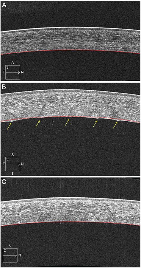
Fig. 1 Image of corneal anterior segment 5-line raster scans from an 80-year-old male before and after uncomplicated phacoemulsification surgery. (A) Preoperative anterior optical coherence tomography (OCT) image. The anterior and posterior surfaces of the cornea had a continuous, smooth contour. (B) Anterior OCT of the cornea one week after surgery. The corneal anterior surface had a continuous, smooth contour, while the posterior corneal surface had 5 points of irregular wave-like appearance (arrows) compared to the posterior virtual contour (red colored dotted lines). (C) Anterior OCT of the cornea one month after surgery. All points of the irregular posterior surface resolved and appeared smooth again. I = inferior; N = nasal; S = superior; T = temporal.
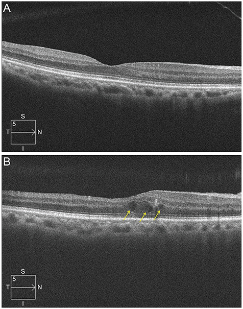
Fig. 2 Macular scan image of the same patient in Fig. 1. (A) Preoperative macular cube scan image. The central subfield retinal thickness (CRT) was 267 µm, and the fovea did not show retinal thickening or cystoid change. (B) Image of macular cube scan 1 month after surgery. CRT increased to 324 µm. The normal foveal depression disappeared, and the images show cystoid change at the fovea (arrows). I = inferior; N = nasal; S = superior; T = temporal.
Reference
-
1. Flach AJ. The incidence, pathogenesis and treatment of cystoid macular edema following cataract surgery. Trans Am Ophthalmol Soc. 1998; 96:557–634.2. Halpern DL, Pasquale LR. Cystoid macular edema in aphakia and pseudophakia after use of prostaglandin analogs. Semin Ophthalmol. 2002; 17:181–186.3. Henderson BA, Kim JY, Ament CS, et al. Clinical pseudophakic cystoid macular edema: risk factors for development and duration after treatment. J Cataract Refract Surg. 2007; 33:1550–1558.4. Loewenstein A, Zur D. Postsurgical cystoid macular edema. Dev Ophthalmol. 2010; 47:148–159.5. Belair ML, Kim SJ, Thorne JE, et al. Incidence of cystoid macular edema after cataract surgery in patients with and without uveitis using optical coherence tomography. Am J Ophthalmol. 2009; 148:128–135.6. Perente I, Utine CA, Ozturker C, et al. Evaluation of macular changes after uncomplicated phacoemulsification surgery by optical coherence tomography. Curr Eye Res. 2007; 32:241–247.7. Benitah NR, Arroyo JG. Pseudophakic cystoid macular edema. Int Ophthalmol Clin. 2010; 50:139–153.8. Setala K. Corneal endothelial cell density in iridocyclitis. Acta Ophthalmol. 1979; 57:277–286.9. Weston BC, Bourne WM, Polse KA, Hodge DO. Corneal hydration control in diabetes mellitus. Invest Ophthalmol Vis Sci. 1995; 36:586–595.10. O'sNeal MR, Polse KA. Decreased endothelial pump function with aging. Invest Ophthalmol Vis Sci. 1986; 27:457–463.11. Glasser DB, Schultz RO, Hyndiuk RA. The role of viscoelastics, cannulas, and irrigating solution additives in post-cataract surgery corneal edema: a brief review. Lens Eye Toxic Res. 1992; 9:351–359.12. Tao A, Chen Z, Shao Y, et al. Phacoemulsification induced transient swelling of corneal Descemet's Endothelium Complex imaged with ultra-high resolution optical coherence tomography. PLoS One. 2013; 8:e80986.13. Behndig A, Lundberg B. Transient corneal edema after phacoemulsification: comparison of 3 viscoelastic regimens. J Cataract Refract Surg. 2002; 28:1551–1556.14. Li YJ, Kim HJ, Joo CK. Early changes in corneal edema following torsional phacoemulsification using anterior segment optical coherence tomography and Scheimpflug photography. Jpn J Ophthalmol. 2011; 55:196–204.15. Zetterstrom C, Laurell CG. Comparison of endothelial cell loss and phacoemulsification energy during endocapsular phacoemulsification surgery. J Cataract Refract Surg. 1995; 21:55–58.16. Hayashi K, Nakao F, Hayashi F. Corneal endothelial cell loss following phacoemulsification using the Small-Port Phaco. Ophthalmic Surg. 1994; 25:510–513.17. Kosrirukvongs P, Slade SG, Berkeley RG. Corneal endothelial changes after divide and conquer versus chip and flip phacoemulsification. J Cataract Refract Surg. 1997; 23:1006–1012.18. Ersoy L, Caramoy A, Ristau T, et al. Aqueous flare is increased in patients with clinically significant cystoid macular oedema after cataract surgery. Br J Ophthalmol. 2013; 97:862–865.19. Shah SM, Spalton DJ. Changes in anterior chamber flare and cells following cataract surgery. Br J Ophthalmol. 1994; 78:91–94.20. Oh JH, Chuck RS, Do JR, Park CY. Vitreous hyper-reflective dots in optical coherence tomography and cystoid macular edema after uneventful phacoemulsification surgery. PLoS One. 2014; 9:e95066.21. Yonekawa Y, Kim IK. Pseudophakic cystoid macular edema. Curr Opin Ophthalmol. 2012; 23:26–32.22. Schmier JK, Halpern MT, Covert DW, Matthews GP. Evaluation of costs for cystoid macular edema among patients after cataract surgery. Retina. 2007; 27:621–628.23. Antcliff RJ, Poulson A, Flanagan DW. Phacoemulsification in diabetics. Eye (Lond). 1996; 10:737–741.24. Morikubo S, Takamura Y, Kubo E, et al. Corneal changes after small-incision cataract surgery in patients with diabetes mellitus. Arch Ophthalmol. 2004; 122:966–969.25. Hariprasad SM, Callanan D, Gainey S, et al. Cystoid and diabetic macular edema treated with nepafenac 0.1%. J Ocul Pharmacol Ther. 2007; 23:585–590.26. Wolf EJ, Braunstein A, Shih C, Braunstein RE. Incidence of visually significant pseudophakic macular edema after uneventful phacoemulsification in patients treated with nepafenac. J Cataract Refract Surg. 2007; 33:1546–1549.27. Miyake K, Ota I, Miyake G, Numaga J. Nepafenac 0.1% versus fluorometholone 0.1% for preventing cystoid macular edema after cataract surgery. J Cataract Refract Surg. 2011; 37:1581–1588.28. Stern AL, Taylor DM, Dalburg LA, Cosentino RT. Pseudophakic cystoid maculopathy: a study of 50 cases. Ophthalmology. 1981; 88:942–946.29. Rossetti L, Autelitano A. Cystoid macular edema following cataract surgery. Curr Opin Ophthalmol. 2000; 11:65–72.30. Shakya K, Pokharel S, Karki KJ, et al. Corneal edema after phacoemulsification surgery in patients with type II diabetes mellitus. Nepal J Ophthalmol. 2013; 5:230–234.
- Full Text Links
-
- Actions
-
Cited
- CITED
-
- Close
- Share
-
- Similar articles
-
- Pathophysiology of Transient Corneal Edema and Pseudophakic Cystoid Macular Edema
- Clinical Results of Nd:Yag Laser Posterior Capsulotomy
- A Case of Secondary Macular Hole Formation after Phacoemulsification in a Vitrectomized Eye
- Prophylactic Intracameral Vancomycin Irrigation and Cystoid Macular Edema
- Assessment for Macular Thickness after Uncomplicated Phacoemulsification Using Optical Coherence Tomography
