Seeding of Meningeal Sarcoma Along a Surgical Trajectory on the Scalp
- Affiliations
-
- 1Department of Neurosurgery, Incheon St. Mary’s Hospital, The Catholic University of Korea, Incheon, Korea. yowas@catholic.ac.kr
- KMID: 2356989
- DOI: http://doi.org/10.14791/btrt.2016.4.2.160
Abstract
- Primary sarcomas of the central nervous system are rare. These tumors is rapid growth often produces mass effect on the brain. Diagnosis is rendered pathologically after resection. Surgical resection is the mainstay treatment and need the adjuvant therapy. We report a 44-year-old female with a meningeal sarcoma of frontal meninges. She complained headache for 2 months and palpable forehead mass for 3 weeks. Brain MRI demonstrated a soft tissue mass sized as 5.3×3.7×3.1 cm with well-defined osteolysis on the midline of the frontal bone. The mass attached to anterior falx without infiltration into the brain parenchyme. The tumor had extracranial and extraaxial extension with bone destruction. The tumor was totally removed with craniectomy and she had an adjuvant radiotherapy. However, an isolated subcutaneous metastasis developed at the both preauricular area of the scalp, originating from the scar which was remained the first surgery. After complete removal of this metastasis, she had an adjuvant radiotherapy in other hospital. However, she expired after six months after first surgery. We believe that the occurrence of tumor seeding at the site of incision in the scalp is related to using the fluid for irrigation after tumor resection and the same surgical instruments for the removal of the brain tumor.
Keyword
MeSH Terms
Figure
-

Fig. 1 Axial CT scan (soft-tissue window) of the head, demonstrating extraaxial and extracranial tumor masses. Axial CT scan (bone window) demonstrating irregular destruction of the skull.
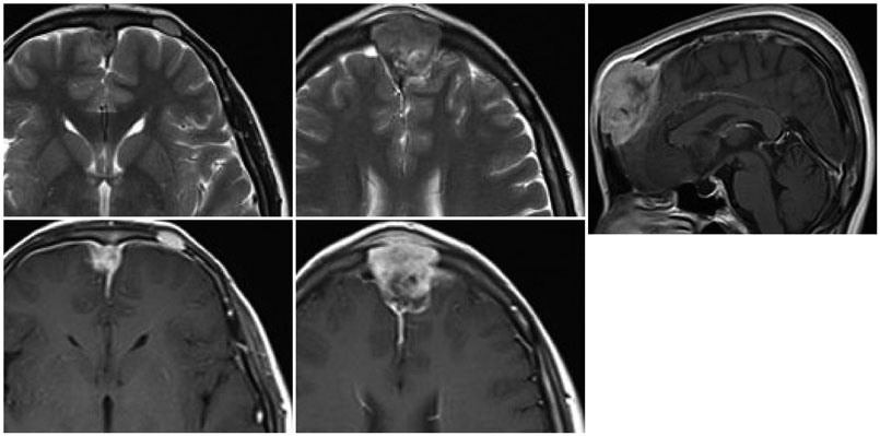
Fig. 2 Axial and sagittal MRI demonstrating an irregular contrast-enhancing mass lesion with compression anterior falx and midline of frontal lobe.
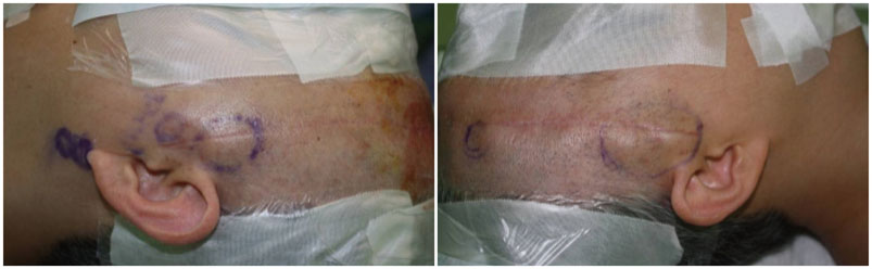
Fig. 3 Gross appearance of the patient’s head with the enlarged, painful palpable mass on the both preauricular area of the scalp along the scar left by the surgery.

Fig. 4 Axial contrast-enhanced MRI of the head demonstrating the heterogeneous contrast-enhancing scalp lesions on the bilateral temporal areas.

Fig. 5 A: Gross appearance of the metastasis at the right preauricular lesion. B: After total removal of the metastatic tumor. C: Gross specimen of removed metastatic tumor.
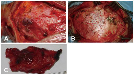
Fig. 6 A: Gross appearance of the metastasis at the left preauricular lesion. B: After total removal of the metastatic tumor. C: Gross specimen of removed metastatic tumor.
Reference
-
1. Palta M, Riedel RF, Vredenburgh JJ, et al. Primary meningeal rhabdomyosarcoma. Sarcoma. 2011; 2011:312802.
Article2. Sugita Y, Shigemori M, Harada H, et al. Primary meningeal sarcomas with leiomyoblastic differentiation: a proposal for a new subtype of primary meningeal sarcomas. Am J Surg Pathol. 2000; 24:1273–1278.3. Cummings M, Chowdhry V, Shah H, Back J, Kennedy GA. Recurrent meningeal sarcoma successfully treated with stereotactic radiosurgery. J Neurosurg Pediatr. 2012; 10:434–438.
Article4. Singla N, Kapoor A, Chatterjee D. Undifferentiated meningeal sarcoma of childhood presenting as hard mass adhered to major intracranial vessels. Childs Nerv Syst. 2016; 32:771–773.
Article5. Maddah G, Shabahang H, Zehi V, Sharifi Sistani N, Mashhadi Nejad H. Iatrogenic seeding of tumor cells in thigh soft tissue upon surgical removal of intracranial meningioma. Basic Clin Neurosci. 2016; 7:159–164.
Article6. Avecillas-Chasin JM, Saceda-Gutierrez J, Alonso-Lera P, et al. Scalp Metastases of Recurrent Meningiomas: Aggressive Behavior or Surgical Seeding? World Neurosurg. 2015; 84:121–131.
Article7. Narayan A, Jallo G, Huisman TA. Extracranial, peritoneal seeding of primary malignant brain tumors through ventriculo-peritoneal shunts in children: case report and review of the literature. Neuroradiol J. 2015; 28:536–539.
Article8. Buis DR, van der Valk P, De Witt Hamer PC. Subcutaneous tumor seeding after biopsy in gliomatosis cerebri. J Neurooncol. 2012; 106:431–435.
Article
