Phantom Study of a New Laser-Etched Needle for Improving Visibility During Ultrasonography-Guided Lumbar Medial Branch Access With Novices
- Affiliations
-
- 1Topteam Rehabilitation Clinic, Gwangju, Korea.
- 2Department of Hospital Biomedical Engineering, Dongshin University, Gwangju, Korea.
- 3Department of Rehabilitation Medicine, Chosun University Hospital, Gwangju, Korea. hayaaaa@hanmail.net
- KMID: 2356642
- DOI: http://doi.org/10.5535/arm.2016.40.4.575
Abstract
OBJECTIVE
To compare the visibility and procedural parameters between a standard spinal needle and a new laser-etched needle (LEN) in real-time ultrasonography guided lumbar medial branch access in a phantom of the lumbosacral spine.
METHODS
We conducted a prospective single-blinded observational study at a rehabilitation medicine center. A new model of LEN was manufactured with a standard 22-gauge spinal needle and a laser etching machine. Thirty-two inexperienced polyclinic medical students performed ultrasonography-guided lumbar medial branch access using both a standard spinal needle and a LEN with scanning protocol. The outcomes included needle visibility score, needle elapsed time, first-pass success rate, and number of needle sticks.
RESULTS
The LEN received significantly better visibility scores and shorter needle elapsed time compared to the standard spinal needle. First-pass success rate and the number of needle sticks were not significantly different between needles.
CONCLUSION
A new LEN is expected to offer better visibility and enable inexperienced users to perform an ultrasonography-guided lumbar medial branch block more quickly. However, further study of variables may be necessary for clinical application.
MeSH Terms
Figure
-
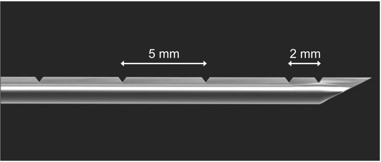
Fig. 1 Computer graphic image of laser-etched needle shows two ultrasound reflectors adjacent to the surface of the needle tip at 2 mm intervals and same-shape ultrasound reflectors located at 5 mm intervals on the same side of the distal needle shaft.
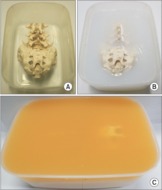
Fig. 2 Gelatin-Metamucil mixture of the lumbosacral spine phantom. (A) An adult-size lumbosacral spine model. (B) Spinal phantom after the lower layer of gelatin mold has set. (C) Yellowish surface layer of the spinal phantom after the Metamucil-mixed gelatin mold has been added.
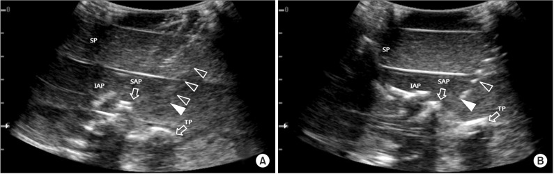
Fig. 3 Ultrasound images of right third lumbar medial branch access with study needles in lumbosacral spine phantom. (A) Using the standard spinal needle. (B) The image of laser-etched needle shows bright dots from the ultrasound reflectors. Open arrowheads, needle shaft; closed arrowheads, needle tip; SP, spinous process; TP, transverse process; IAP, inferior articular process; SAP, superior articular process.
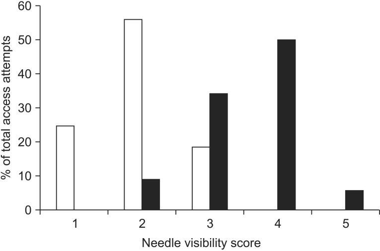
Fig. 4 Estimates of needle visibility for standard spinal needle (gray bars) and laser-etched needle (black bars).
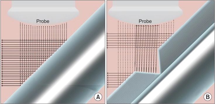
Fig. 5 Schematic diagram of ultrasound performance with the inplane approach. (A) Ultrasound beam is reflected off of standard spinal needle surface away from probe. (B) Due to wedge shaped reflectors, part of the ultrasound beam is reflected back to the probe.
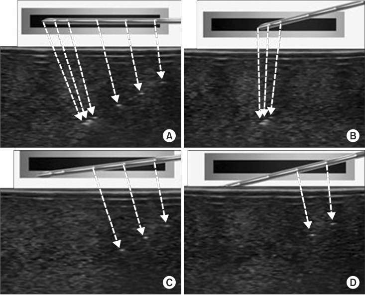
Fig. 6 Ultrasound images of laser-etched needle by location and direction of ultrasound beam (black bar) in soft tissue phantom. Dotted arrows, reflectors of laser-etched needle.
Reference
-
1. Greher M, Scharbert G, Kamolz LP, Beck H, Gustorff B, Kirchmair L, et al. Ultrasound-guided lumbar facet nerve block: a sonoanatomic study of a new methodologic approach. Anesthesiology. 2004; 100:1242–1248. PMID: 15114223.2. Saal JS. General principles of diagnostic testing as related to painful lumbar spine disorders: a critical appraisal of current diagnostic techniques. Spine (Phila Pa 1976). 2002; 27:2538–2545. PMID: 12435989.3. Ha DH, Shim DM, Kim TK, Kim YM, Choi SS. Comparison of ultrasonography- and fluoroscopy-guided facet joint block in the lumbar spine. Asian Spine J. 2010; 4:15–22. PMID: 20622950.
Article4. Kaplan M, Dreyfuss P, Halbrook B, Bogduk N. The ability of lumbar medial branch blocks to anesthetize the zygapophysial joint: a physiologic challenge. Spine (Phila Pa 1976). 1998; 23:1847–1852. PMID: 9762741.5. Chin KJ, Perlas A, Chan VW, Brull R. Needle visualization in ultrasound-guided regional anesthesia: challenges and solutions. Reg Anesth Pain Med. 2008; 33:532–544. PMID: 19258968.
Article6. Crum T, Adhikari S, Lander L, Blaivas M. Do echo-enhanced needles make a difference in sonographically guided vascular access? J Ultrasound Med. 2014; 33:623–628. PMID: 24658941.
Article7. Kilicaslan A, Topal A, Tavlan A, Erol A, Otelcioglu S. Differences in tip visibility and nerve block parameters between two echogenic needles during a simulation study with inexperienced anesthesia trainees. J Anesth. 2014; 28:460–462. PMID: 24127134.
Article8. Marks R, Semple AJ. Spinal anaesthesia after facet joint injection. Anaesthesia. 1988; 43:65–66. PMID: 3344957.
Article9. Sites BD, Spence BC, Gallagher JD, Wiley CW, Bertrand ML, Blike GT. Characterizing novice behavior associated with learning ultrasound-guided peripheral regional anesthesia. Reg Anesth Pain Med. 2007; 32:107–115. PMID: 17350520.
Article10. Gottlieb RH, Robinette WB, Rubens DJ, Hartley DF, Fultz PJ, Violante MR. Coating agent permits improved visualization of biopsy needles during sonography. AJR Am J Roentgenol. 1998; 171:1301–1302. PMID: 9798867.
Article11. Culp WC, McCowan TC, Goertzen TC, Habbe TG, Hummel MM, LeVeen RF, et al. Relative ultrasonographic echogenicity of standard, dimpled, and polymeric-coated needles. J Vasc Interv Radiol. 2000; 11:351–358. PMID: 10735431.
Article12. Barr RG. Improved needle visualization with electronic beam steering: proof of concept. Ultrasound Q. 2012; 28:59–64. PMID: 22634767.13. Hocking G, Mitchell CH. Optimizing the safety and practice of ultrasound-guided regional anesthesia: the role of echogenic technology. Curr Opin Anaesthesiol. 2012; 25:603–609. PMID: 22825047.14. Edgcombe H, Hocking G. Sonographic identification of needle tip by specialists and novices: a blinded comparison of 5 regional block needles in fresh human cadavers. Reg Anesth Pain Med. 2010; 35:207–211. PMID: 20301826.15. Uppal V, Sondekoppam RV, Ganapathy S. Effect of beam steering on the visibility of echogenic and non-echogenic needles: a laboratory study. Can J Anaesth. 2014; 61:909–915. PMID: 25053210.
Article16. Bellingham GA, Peng PW. A low-cost ultrasound phantom of the lumbosacral spine. Reg Anesth Pain Med. 2010; 35:290–293. PMID: 20921841.
Article17. Kendall JL, Faragher JP. Ultrasound-guided central venous access: a homemade phantom for simulation. CJEM. 2007; 9:371–373. PMID: 17935654.
Article18. Greher M, Kirchmair L, Enna B, Kovacs P, Gustorff B, Kapral S, et al. Ultrasound-guided lumbar facet nerve block: accuracy of a new technique confirmed by computed tomography. Anesthesiology. 2004; 101:1195–1200. PMID: 15505456.19. Guo S, Schwab A, McLeod G, Corner G, Cochran S, Eisma R, et al. Echogenic regional anaesthesia needles: a comparison study in Thiel cadavers. Ultrasound Med Biol. 2012; 38:702–707. PMID: 22390992.
Article20. Rauch S, Kasuya Y, Turan A, Neamtu A, Vinayakan A, Sessler DI. Ultrasound-guided lumbar medial branch block in obese patients: a fluoroscopically confirmed clinical feasibility study. Reg Anesth Pain Med. 2009; 34:340–342. PMID: 19585701.21. Miura M, Takeyama K, Suzuki T. Visibility of ultrasound-guided echogenic needle and its potential in clinical delivery of regional anesthesia. Tokai J Exp Clin Med. 2014; 39:80–86. PMID: 25027252.22. Sviggum HP, Ahn K, Dilger JA, Smith HM. Needle echogenicity in sonographically guided regional anesthesia: blinded comparison of 4 enhanced needles and validation of visual criteria for evaluation. J Ultrasound Med. 2013; 32:143–148. PMID: 23269719.23. Gofeld M, Krashin DL, Ahn S. Needle echogenicity in ultrasound-guided lumbar spine injections: a cadaveric study. Pain Physician. 2013; 16:E725–E730. PMID: 24284853.24. Deam RK, Kluger R, Barrington MJ, McCutcheon CA. Investigation of a new echogenic needle for use with ultrasound peripheral nerve blocks. Anaesth Intensive Care. 2007; 35:582–586. PMID: 18020079.
Article25. Maecken T, Zenz M, Grau T. Ultrasound characteristics of needles for regional anesthesia. Reg Anesth Pain Med. 2007; 32:440–447. PMID: 17961844.
Article26. Schafhalter-Zoppoth I, McCulloch CE, Gray AT. Ultrasound visibility of needles used for regional nerve block: an in vitro study. Reg Anesth Pain Med. 2004; 29:480–488. PMID: 15372394.27. Nichols K, Wright LB, Spencer T, Culp WC. Changes in ultrasonographic echogenicity and visibility of needles with changes in angles of insonation. J Vasc Interv Radiol. 2003; 14:1553–1557. PMID: 14654490.
Article
- Full Text Links
-
- Actions
-
Cited
- CITED
-
- Close
- Share
-
- Similar articles
-
- Ultrasound Phantoms to Protect Patients from Novices
- Burn Wound along the Guide Needle Trajectory as a Complication of Radiofrequency Neurotomy of the Lumbar Medial Branch: A case report
- Transverse Process and Needles of Medial Branch Block to Facet Joint as Landmarks for Ultrasound-Guided Selective Nerve Root Block
- Measuring Needle Angle and Depth for Lumbar Medial Branch Block Using Ultrasonography: An Evaluation of Efficiency Compared with Magnetic Resonance Imaging
- Comparison of Lidocaine and Bupivacaine in Lumbar Medial Branch Block
