Radix mesiolingualis and radix distolingualis: a case report of a tooth with an unusual morphology
- Affiliations
-
- 1Department of Conservative Dentistry and Endodontics, Seema Dental College and Hospital, Rishikesh, Uttarakhand, India. gurudutt_nayak@hotmail.com
- 2Department of Prosthodontics, Seema Dental College and Hospital, Rishikesh, Uttarakhand, India.
- 3Dasmesh Institute of Research and Dental Science, Faridkot, Punjab, India.
- KMID: 2356010
- DOI: http://doi.org/10.5395/rde.2016.41.4.322
Abstract
- Variation in the root and canal morphology of the maxillary first molars is quite common. The most common configuration is 3 roots and 3 or 4 canals. Nonetheless, other possibilities still exist. The presence of an additional palatal root is rather uncommon and has been reported to have an incidence of 0.06 - 1.6% in varying populations studied. Whenever two palatal roots exist, one of them is the normal palatal root, the other is a supernumerary structure which can be located either mesiolingually (radix mesiolingualis) or distolingually (radix distolingualis). This case report describes successful endodontic treatment of a maxillary first molar with radix mesiolingualis and radix distolingualis. Identification of this variation was done through clinical examination along with the aid of multiangled radiographs, and an accurate assessment of this morphology was made with the help of a cone-beam computed tomography imaging. In addition to the literature review, this article also discusses the epidemiology, classifications, morphometric features, guidelines for diagnosis, and endodontic management of a maxillary first molar with extra-palatal root.
Keyword
Figure
-
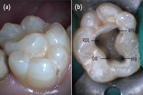
Figure 1 (a) Palatal surface of tooth #16 showing three well-developed lobulated cusps; (b) Access opening showing four root canal orifices of tooth #16. MB, mesiobuccal; DB, distobuccal; RML, radix mesiolingualis; RDL, radix distolingualis.
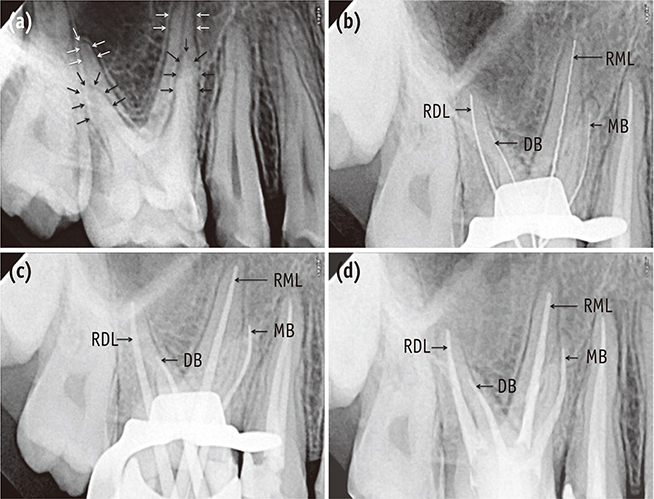
Figure 2 (a) A preoperative radiograph of teeth #15 and 16 (white arrows showing the outlines of the palatal roots and black arrows showing the outlines of the buccal roots of tooth #16); (b) Working length radiographs of tooth #16; (c) Master cone radiograph of tooth #16; (d) Post-obturation radiograph of tooth #16. MB, mesiobuccal; DB, distobuccal; RML, radix mesiolingualis; RDL, radix distolingualis.
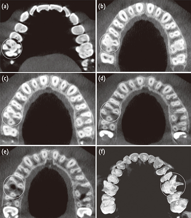
Figure 3 Axial section of cone-beam computed tomography (CBCT) scan images of tooth #16 (a - e). (a) Showing three cusps on the palatal surface formed of enamel overgrowth; (b) Showing floor of the pulp chamber; (c) Showing two buccal and two palatal roots at the cervical third; (d) At the middle; (e) At the apical third level (rounded area). (f) A 3 dimensional reconstruction image of the maxilla showing two buccal and two palatal roots (rounded area).

Figure 4 Cone-beam computed tomography (CBCT) scan images of tooth #16. (a) Transverse section scan image showing the angle of divergence between the two palatal roots; (b - e) Axial sections; (b) Showing the distance between the palatal root canal orifices; (c) The distance between the buccal root canal orifices; (d) The distopalatal angle; (e) Mesiopalatal angle.
Reference
-
1. Ree M, Schwartz RS. The endo-restorative interface: current concepts. Dent Clin North Am. 2010; 54:345–374.
Article2. Burns RC, Buchanan LS. Tooth morphology and access openings. In : Cohen S, Burns RC, editors. Pathways of the pulp. 6th ed. St Louis: Mosby;1987. p. 148.3. Gopikrishna V, Bhargavi N, Kandaswamy D. Endodontic management of a maxillary first molar with a single root and a single canal diagnosed with the aid of spiral CT: a case report. J Endod. 2006; 32:687–691.
Article4. Nayak G, Dahiya S, Singh I, Mohammad FH. Endodontic management of an unusual maxillary first molar with a single buccal root. J Contemp Dent Pract. 2014; 15:367–371.
Article5. Adanir N. An unusual maxillary first molar with four roots and six canals: a case report. Aust Dent J. 2007; 52:333–335.
Article6. Barbizam JV, Ribeiro RG, Tanomaru Filho M. Unusual anatomy of permanent maxillary molars. J Endod. 2004; 30:668–671.
Article7. Kottoor J, Velmurugan N, Surendran S. Endodontic management of a maxillary first molar with eight root canal systems evaluated using cone-beam computed tomography scanning: a case report. J Endod. 2011; 37:715–719.
Article8. Carlsen O, Alexandersen V. Radix mesiolingualis and radix distolingualis in a collection of permanent maxillary molars. Acta Odontol Scand. 2000; 58:229–236.
Article9. Carlsen O, Alexandersen V. Radix paramolaris and radix distomolaris in Danish permanent maxillary molars. Acta Odontol Scand. 1999; 57:283–289.
Article10. Kottoor J, Nandini S, Velmurugan N. Maxillary first molar with three buccal roots evaluated with cone-beam computed tomography: a rare case report. Gen Dent. 2012; 60:e404–e407.11. Rajalbandi S, Shingte SN, Sundaresh KJ, Mallikarjuna R. Aberration in the palatal root of the maxillary first molar. BMJ Case Rep. 2013; 04. 30. DOI: 10.1136/bcr-2013-008641. [Epub ahead of print].
Article12. Kottoor J, Velmurugan N, Ballal S, Roy A. Four-rooted maxillary first molar having C-shaped palatal root canal morphology evaluated using cone-beam computerized tomography: a case report. Oral Surg Oral Med Oral Pathol Oral Radiol Endod. 2011; 111:e41–e45.
Article13. He W, Wei K, Chen J, Yu Q. Endodontic treatment of maxillary first molars presenting with unusual asymmetric palatal root morphology using spiral computerized tomography: a case report. Oral Surg Oral Med Oral Pathol Oral Radiol Endod. 2010; 109:e55–e59.
Article14. Tomazinho FS, Baratto-Filho F, Zaitter S, Leonardi DP, Gonzaga CC. Unusual anatomy of a maxillary first molar with two palatal roots: a case report. J Oral Sci. 2010; 52:149–153.
Article15. Chakradhar Raju RV, Chandrasekhar V, Singh CV, Pasari S. Maxillary molar with two palatal roots: two case reports. J Conserv Dent. 2010; 13:58–61.
Article16. Holderrieth S, Gernhardt CR. Maxillary molars with morphologic variations of the palatal root canals: a report of four cases. J Endod. 2009; 35:1060–1065.
Article17. Gopikrishna V, Reuben J, Kandaswamy D. Endodontic management of a maxillary first molar with two palatal roots and a single fused buccal root diagnosed with spiral computed tomography - a case report. Oral Surg Oral Med Oral Pathol Oral Radiol Endod. 2008; 105:e74–e78.
Article18. Baratto-Filho F, Fariniuk LF, Ferreira EL, Pecora JD, Cruz-Filho AM, Sousa-Neto MD. Clinical and macroscopic study of maxillary molars with two palatal roots. Int Endod J. 2002; 35:796–801.
Article19. Christie WH, Peikoff MD, Fogel HM. Maxillary molars with two palatal roots: a retrospective clinical study. J Endod. 1991; 17:80–84.
Article20. Di Fiore PM. A four-rooted quadrangular maxillary molar. J Endod. 1999; 25:695–697.
Article21. Thews ME, Kemp WB, Jones CR. Aberrations in palatal root and root canal morphology of two maxillary first molars. J Endod. 1979; 5:94–96.
Article22. Türp JC, Alt KW. Chapter 3.1. Anatomy and morphology of human teeth. In : Alt KW, Rösing FW, Teschler-Nicola M, editors. Dental anthropology. Fundamentals, limits and prospects. Wien: Springer-Verlag;1998. p. 71–94.23. Cleghorn BM, Christie WH, Dong CC. Root and root canal morphology of the human permanent maxillary first molar: a literature review. J Endod. 2006; 32:813–821.
Article24. Neelakantan P, Subbarao C, Ahuja R, Subbarao CV, Gutmann JL. Cone-beam computed tomography study of root and canal morphology of maxillary first and second molars in an Indian population. J Endod. 2010; 36:1622–1627.
Article25. Yang B, Lu Q, Bai QX, Zhang Y, Liu XJ, Liu ZJ. Evaluation of the prevalence of the maxillary molars with two palatal roots by cone-beam CT. Zhonghua Kou Qiang Yi Xue Za Zhi. 2013; 48:359–362.26. Rouhani A, Bagherpour A, Akbari M, Azizi M, Nejat A, Naghavi N. Cone-beam computed tomography evaluation of maxillary first and second molars in Iranian population: a morphological study. Iran Endod J. 2014; 9:190–194.27. Gu Y, Wang W, Ni L. Four-rooted permanent maxillary first and second molars in a northwestern Chinese population. Arch Oral Biol. 2015; 60:811–817.
Article28. Nikoloudaki GE, Kontogiannis TG, Kerezoudis NP. Evaluation of the root and canal morphology of maxillary permanent molars and the incidence of the second mesiobuccal root canal in Greek population using cone-beam computed tomography. Open Dent J. 2015; 31:267–272.
Article29. Tian XM, Yang XW, Qian L, Wei B, Gong Y. Analysis of the root and canal morphologies in maxillary first and second molars in a Chinese population using cone-beam computed tomography. J Endod. 2016; 42:696–701.
Article30. Diamond M. Dental anatomy including anatomy of head and neck. 3rd ed. New York: MacMillan;1952. p. 203–205.31. Versiani MA, Pécora JD, de Sousa-Neto MD. Root and root canal morphology of four-rooted maxillary second molars: a micro-computed tomography study. J Endod. 2012; 38:977–982.
Article32. Sabala CL, Benenati FW, Neas BR. Bilateral root or root canal aberrations in a dental school patient population. J Endod. 1994; 20:38–42.
Article33. Benenati FW. Maxillary second molar with two palatal canals and a palatogingival groove. J Endod. 1985; 11:308–310.
Article34. Calberson FL, de Moor RJ, Deroose CA. The radix entomolaris and paramolaris: clinical approach in endodontics. J Endod. 2007; 33:58–63.
Article35. Kannan SK, Suganya , Santharam H. Supernumerary roots. Indian J Dent Res. 2002; 13:116–119.36. Ahmed HM, Abbott PV. Accessory roots in maxillary molar teeth: a review and endodontic considerations. Aust Dent J. 2012; 57:123–131.
Article
- Full Text Links
-
- Actions
-
Cited
- CITED
-
- Close
- Share
-
- Similar articles
-
- Radix augmentation using temporalis fascia graft
- Asymmetry in mesial root number and morphology in mandibular second molars: a case report
- Effects of Polygalae Radix on Apomorphine-Induced Stereotyped Behaviors in Mice
- Effects of Dichloromethane Fraction of Phlomidis Radix on Bone Formation in Human Fetal Osteoblasts
- Effects of Ethanolic Extracts of Scutellaria Radix on the alveolar bone formation in the extract socket of rat
