Microsurgical re-treatment of an endodontically treated tooth with an apically located incomplete vertical root fracture: a clinical case report
- Affiliations
-
- 1Università degli Studi di Milano, Dipartimento di Scienze Biomediche, Chirurgiche e Odontoiatriche, IRCCS Istituto Ortopedico Galeazzi, Milano, Italy. stefano.corbella@gmail.com
- 2Conservative Dentistry and Endodontic Department, Faculty of Dentistry, Mansoura University, Mansoura, Egypt.
- 3Department of Endodontology, Maurice and Gabriela Goldschleger School of Dental Medicine, Tel Aviv University, Tel Aviv, Israel.
- KMID: 2356009
- DOI: http://doi.org/10.5395/rde.2016.41.4.316
Abstract
- Although it is challenging, the early diagnosis of a vertical root fracture (VRF) is crucial in order to ensure tooth preservation. The purpose of this clinical case report was to describe reparative surgery performed to treat a tooth affected by an incomplete VRF. A 26 year old male patient was suspected to have a VRF in a maxillary left central incisor, and an exploratory flap was performed in order to confirm the diagnosis. After detecting the fracture, the lesion was surgically treated, the fracture and the infected root-end were removed, and a platelet-rich plasma membrane was used to cover the defect in order to prevent bacterial migration. A 24 month clinical and radiological follow-up examination showed that the tooth was asymptomatic and that the healing process was in progress. The surgical approach described here may be considered an effective treatment for a combined endodontic-periodontal lesion originating from an incomplete VRF and a recurrent periapical lesion.
MeSH Terms
Figure
-
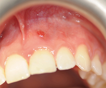
Figure 1 Clinical presentation at baseline. The presence of a sinus tract is evident.
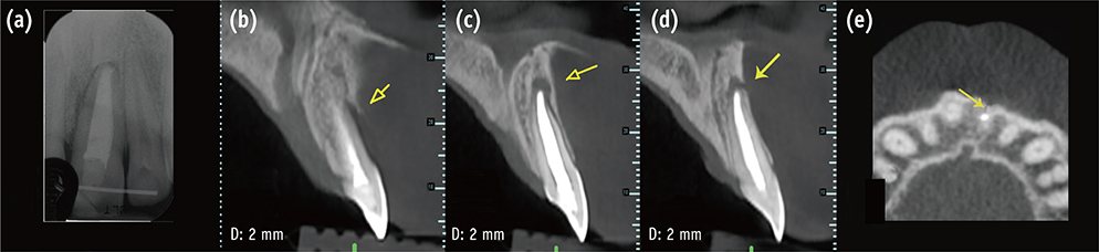
Figure 2 (a) Periapical radiograph and (b, c, d, and e) cone-beam computed tomography (CBCT) sections showing the presence of a periradicular lesion. (b, c, and d) The sagittal sections show the periapical bone defect; (e) the CBCT horizontal projection demonstrates the fracture line.
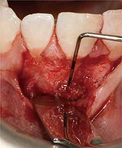
Figure 3 After flap elevation, the presence of the lesion was clearly detectable.
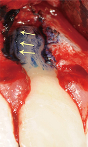
Figure 4 Visualization of the vertical root fracture on the root.
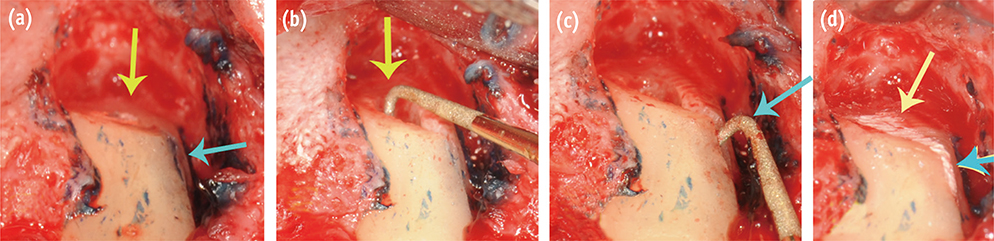
Figure 5 (a) After root resection, the root canal orifice is clearly visualized (yellow), as well as the small residual fracture line (blue); (b) The root-end preparation using a piezoelectric device is shown; (c) Groove preparation following the residual fracture line; (d) Filling of the created cavity.
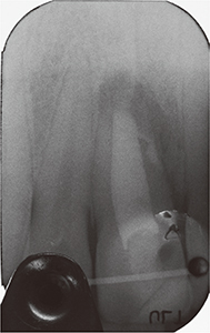
Figure 6 Radiograph taken immediately after surgery.
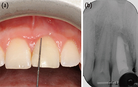
Figure 7 Results of clinical (a) and radiographic (b) examinations six months after surgery.
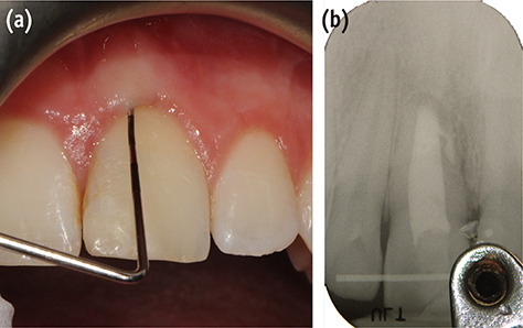
Figure 8 Results of clinical (a) and radiographic (b) examinations 24 months after surgery.
Reference
-
1. Rivera EM, Walton RE. Longitudinal tooth fractures: findings that contribute to complex endodontic diagnoses. Endod Topics. 2007; 16:82–111.
Article2. Lam PP, Palamara JE, Messer HH. Fracture strength of tooth roots following canal preparation by hand and rotary instrumentation. J Endod. 2005; 31:529–532.
Article3. Corbella S, Taschieri S, Samaranayake L, Tsesis I, Nemcovsky C, Del Fabbro M. Implant treatment choice after extraction of a vertically fractured tooth. A proposal for a clinical classification of bony defects based on a systematic review of literature. Clin Oral Implants Res. 2014; 25:946–956.
Article4. Tsesis I, Rosen E, Tamse A, Taschieri S, Kfir A. Diagnosis of vertical root fractures in endodontically treated teeth based on clinical and radiographic indices: a systematic review. J Endod. 2010; 36:1455–1458.
Article5. Tamse A, Fuss Z, Lustig J, Ganor Y, Kaffe I. Radiographic features of vertically fractured, endodontically treated maxillary premolars. Oral Surg Oral Med Oral Pathol Oral Radiol Endod. 1999; 88:348–352.
Article6. Tamse A, Kaffe I, Lustig J, Ganor Y, Fuss Z. Radiographic features of vertically fractured endodontically treated mesial roots of mandibular molars. Oral Surg Oral Med Oral Pathol Oral Radiol Endod. 2006; 101:797–802.
Article7. Corbella S, Del Fabbro M, Tamse A, Rosen E, Tsesis I, Taschieri S. Cone beam computed tomography for the diagnosis of vertical root fractures: a systematic review of the literature and meta-analysis. Oral Surg Oral Med Taschieri S et al. Oral Surg Oral Med Oral Pathol Oral Radiol. 2014; 118:593–602.
Article8. Talwar S, Utneja S, Nawal RR, Kaushik A, Srivastava D, Oberoy SS. Role of cone-beam computed tomography in diagnosis of vertical root fractures: a systematic review and meta-analysis. J Endod. 2016; 42:12–24.
Article9. Chang E, Lam E, Shah P, Azarpazhooh A. Cone-beam computed tomography for detecting vertical root fractures in endodontically treated teeth: a systematic review. J Endod. 2016; 42:177–185.
Article10. Pitts DL, Natkin E. Diagnosis and treatment of vertical root fractures. J Endod. 1983; 9:338–346.
Article11. Taschieri S, Tamse A, Del Fabbro M, Rosano G, Tsesis I. A new surgical technique for preservation of endodontically treated teeth with coronally located vertical root fractures: a prospective case series. Oral Surg Oral Med Oral Pathol Oral Radiol Endod. 2010; 110:e45–e52.
Article12. Moule AJ, Kahler B. Diagnosis and management of teeth with vertical root fractures. Aust Dent J. 1999; 44:75–87.
Article13. Kawai K, Masaka N. Vertical root fracture treated by bonding fragments and rotational replantation. Dent Traumatol. 2002; 18:42–45.
Article14. Velvart P, Ebner-Zimmermann U, Ebner JP. Comparison of papilla healing following sulcular full-thickness flap and papilla base flap in endodontic surgery. Int Endod J. 2003; 36:653–659.
Article15. Velvart P, Ebner-Zimmermann U, Ebner JP. Comparison of long-term papilla healing following sulcular full thickness flap and papilla base flap in endodontic surgery. Int Endod J. 2004; 37:687–693.
Article16. Del Fabbro M, Tsesis I, Rosano G, Bortolin M, Taschieri S. Scanning electron microscopic analysis of the integrity of the root-end surface after root-end management using a piezoelectric device: a cadaveric study. J Endod. 2010; 36:1693–1697.
Article17. Lustig JP, Tamse A, Fuss Z. Pattern of bone resorption in vertically fractured, endodontically treated teeth. Oral Surg Oral Med Oral Pathol Oral Radiol Endod. 2000; 90:224–227.
Article18. Corbella S, Taschieri S, Elkabbany A, Del Fabbro M, von Arx T. Guided tissue regeneration using a barrier membrane in endodontic surgery. Swiss Dent J. 2016; 126:13–25.19. von Arx T, Cochran DL. Rationale for the application of the GTR principle using a barrier membrane in endodontic surgery: a proposal of classification and literature review. Int J Periodontics Restorative Dent. 2001; 21:127–139.20. Gagliani MM, Gorni FG, Strohmenger L. Periapical resurgery versus periapical surgery: a 5-year longitudinal comparison. Int Endod J. 2005; 38:320–327.
Article21. Peterson J, Gutmann JL. The outcome of endodontic resurgery: a systematic review. Int Endod J. 2001; 34:169–175.
Article22. Del Fabbro M, Ceresoli V, Lolato A, Taschieri S. Effect of platelet concentrate on quality of life after periradicular surgery: a randomized clinical study. J Endod. 2012; 38:733–739.
Article23. Taschieri S, Corbella S, Tsesis I, Del Fabbro M. Impact of the use of plasma rich in growth factors (PRGF) on the quality of life of patients treated with endodontic surgery when a perforation of sinus membrane occurred. A comparative study. Oral Maxillofac Surg. 2014; 18:43–52.
Article24. Anitua E, Murias-Freijo A, Alkhraisat MH, Orive G. Clinical, radiographical, and histological outcomes of plasma rich in growth factors in extraction socket: a randomized controlled clinical trial. Clin Oral Investig. 2015; 19:589–600.
Article25. Fabbro MD, Bortolin M, Taschieri S, Ceci C, Weinstein RL. Antimicrobial properties of platelet-rich preparations. A systematic review of the current preclinical evidence. Platelets. 2016; 27:276–285.
Article26. Drago L, Bortolin M, Vassena C, Romanò CL, Taschieri S, Del Fabbro M. Plasma components and platelet activation are essential for the antimicrobial properties of autologous platelet-rich plasma: an in vitro study. PLoS One. 2014; 9:e107813.
- Full Text Links
-
- Actions
-
Cited
- CITED
-
- Close
- Share
-
- Similar articles
-
- Influence of various properties of post and core on the stress distribution in endodontically treated tooth
- Post and core build-ups in crown and bridge abutments: Bio-mechanical advantages and disadvantages
- Healing after horizontal root fractures: 3 cases with 2-year follow-up
- Factors Affecting the Pulp and Root Healing of Root Fractures in Immature Permanent Teeth
- Critical evaluation of fracture strength testing for endodontically treated teeth: a finite element analysis study
