Native T1 Mapping Demonstrating Apical Thrombi in Eosinophilic Myocarditis Associated with Churg-Strauss Syndrome
- Affiliations
-
- 1Department of Radiology, Seoul St. Mary's Hospital, College of Medicine, The Catholic University, Seoul, Korea. jijung@catholic.ac.kr
- 2Department of Radiology, Yeouido St. Mary's Hospital, College of Medicine, The Catholic University, Seoul, Korea.
- 3Department of Pathology, Seoul St. Mary's Hospital, College of Medicine, The Catholic University, Seoul, Korea.
- 4Division of Cardiology, Department of Internal Medicine, Seoul St. Mary's Hospital, College of Medicine, The Catholic University, Seoul, Korea.
- KMID: 2355469
- DOI: http://doi.org/10.4070/kcj.2016.46.6.882
Abstract
- Eosinophilic myocarditis is a disease characterized by eosinophilic infiltration of the myocardium, consisting of acute necrotic stage, thrombotic stage, and fibrotic stage. Although T1 mapping has been increasingly used in various cardiac pathologies, there has been no report of T1 mapping in eosinophilic myocarditis. We report a case of 75-year-old female with eosinophilic myocarditis, whose cardiac magnetic resonance imaging included native T1 mapping, in which apical thrombi were distinctly seen as areas with decreased T1 values, next to areas of inflammation seen as increased T1 value in subendocardium.
MeSH Terms
Figure
-
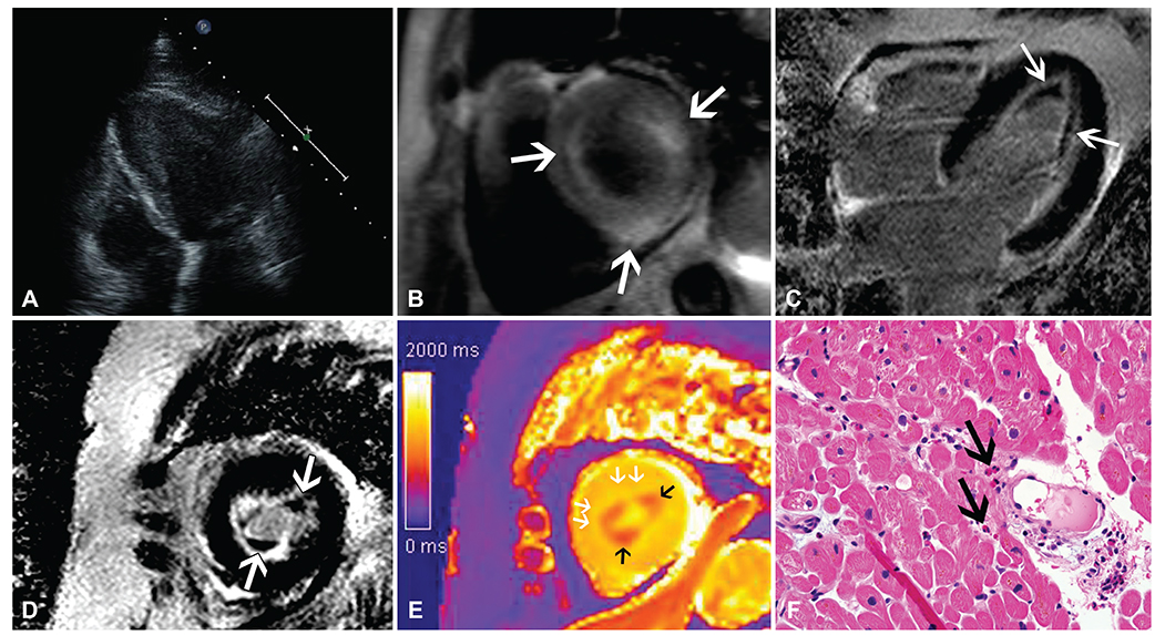
Fig. 1 Eosinophilic myocarditis associated with Churg-Strauss syndrome in a 75-year-old woman. (A) Apical four-chamber view of the heart on echocardiogram shows hypertrophied left ventricular apex with impaired relaxation. (B) T2-weighted image of the heart, short axis view demonstrates multifocal, patchy high signal intensity lesions (arrows) throughout the left ventricular wall, suggesting areas of edema. (C, D) Late Gadolinium enhancement (LGE) images in four-chamber view (C) and short axis view (D) demonstrate diffuse subendocardial enhancement in the left ventricular wall with non-enhancing thrombi (arrows) in the apex. (E) Cardiac MR mapping images of the heart. T1 mapping of the apex demonstrates oval areas (black arrows) with decreased T1 value (1007±84.67 ms), which correspond to the mural thrombi seen on LGE images. T1 value of subendocardium immediately adjacent to the mural thrombi (white arrows) are diffusely increased (1527±67.41 ms), corresponding to areas that show subendocardial enhancement on LGE images (reference value: 1278±30 ms). (F) Myocardial biopsy specimen shows infiltration of eosinophils (arrows) in the myocardium, confirming the diagnosis of eosinophilic myocarditis (H-E, x400).
Reference
-
1. Ugander M, Oki AJ, Hsu LY, et al. Extracellular volume imaging by magnetic resonance imaging provides insights into overt and sub-clinical myocardial pathology. Eur Heart J. 2012; 33:1268–1278.2. Masi AT, Hunder GG, Lie JT, et al. The American College of Rheumatology 1990 criteria for the classification of Churg-Strauss syndrome (allergic granulomatosis and angiitis). Arthritis Rheum. 1990; 33:1094–1100.3. Thambidorai SK, Korlakunta HL, Arouni AJ, Hunter WJ, Holmberg MJ. Acute eosinophilic myocarditis mimicking myocardial infarction. Tex Heart Inst J. 2009; 36:355–357.4. Olsen EG. Restrictive cardiomyopathy. Postgrad Med J. 1986; 62:607–608.5. Yoon YE, Hong YJ, Kim HK, et al. 2014 Korean guidelines for appropriate utilization of cardiovascular magnetic resonance imaging: a joint report of the korean society of cardiology and the korean society of radiology. Korean Circ J. 2014; 44:359–385.6. Tani H, Amano Y, Tachi M, Machida T, Mizuno K, Kumita S. T2-weighted and delayed enhancement MRI of eosinophilic myocarditis: relationship with clinical phases and global cardiac function. Jpn J Radiol. 2012; 30:824–831.7. Gupta A, Singh Gulati G, Seth S, Sharma S. Cardiac MRI in restrictive cardiomyopathy. Clin Radiol. 2012; 67:95–105.8. Salanitri GC. Endomyocardial fibrosis and intracardiac thrombus occurring in idiopathic hypereosinophilic syndrome. AJR Am J Roentgenol. 2005; 184:1432–1433.9. Chang SA, Kim HK, Park EA, Kim YJ, Sohn DW. Images in cardiovascular medicine. Loeffler endocarditis mimicking apical hypertrophic cardiomyopathy. Circulation. 2009; 120:82–85.10. Debl K, Djavidani B, Buchner S, et al. Time course of eosinophilic myocarditis visualized by CMR. J Cardiovasc Magn Reson. 2008; 10:21.11. Hamlin SA, Henry TS, Little BP, Lerakis S, Stillman AE. Mapping the future of cardiac MR imaging: case-based review of T1 and T2 mapping techniques. Radiographics. 2014; 34:1594–1611.12. Karamitsos TD, Piechnik SK, Banypersad SM, et al. Noncontrast T1 mapping for the diagnosis of cardiac amyloidosis. JACC Cardiovasc Imaging. 2013; 6:488–497.
- Full Text Links
-
- Actions
-
Cited
- CITED
-
- Close
- Share
-
- Similar articles
-
- Eosinophilic Annular Erythema in a Patient with Eosinophilic Granulomatosis with Polyangiitis (Churg-Strauss Syndrome)
- A Case of Churg-Strauss Syndrome with Endomyocardial Fibrosis
- An atypical case of Churg-Strauss syndrome without asthma
- A Case of Loeffler's Endocarditis Associated with Churg-Strauss Syndrome
- A case of cardiac involvement in Churg-Strauss syndrome presenting without cardiomegaly in an asymptomatic patient
