En Plaque Tuberculoma: a Case Report
- Affiliations
-
- 1Department of Radiology, Seoul Medical Center, Seoul, Korea. jnoon276@gmail.com
- KMID: 2354803
- DOI: http://doi.org/10.13104/imri.2016.20.3.200
Abstract
- In Korea, tuberculosis is still common disease. Central nervous system tuberculosis can manifest in a variety of forms, including tuberculous meningitis, tuberculous cerebritis, tuberculoma, tuberculous abscess, and miliary tuberculosis. Although intra-axial tuberculomas are the more common type of CNS tuberculosis, extra-axial lesions are rarely encountered. En plaque tuberculoma is an extremely rare presentation of intracranial tuberculosis with mimicking primary or secondary meningeal neoplasia. We describe a rare case of an en plaque tuberculoma accompanied by tuberculous meningitis and tuberculomas.
MeSH Terms
Figure
-

Fig. 1 A 73-year-old woman with an en plaque tuberculoma. (a) CT scan showed iso to hyperattenuating lesion at left temporal extra-axial space with low density area in the Lt. temporal lobe. (b) DWI showed iso to low signal intensity lesion at left temporal extra-axial space. (c) T2-weighted axial MRI showed iso-signal intensity lesion with perilesional high signal intensity. (d-f) T1-weighted axial (d) MRI showed iso-signal intensity lesion with contrast-enhanced T1-weighted axial (e) and coronal (f) MRI showing homogeneous enhancing dural-based lesion in the left temporal convexity (arrows).
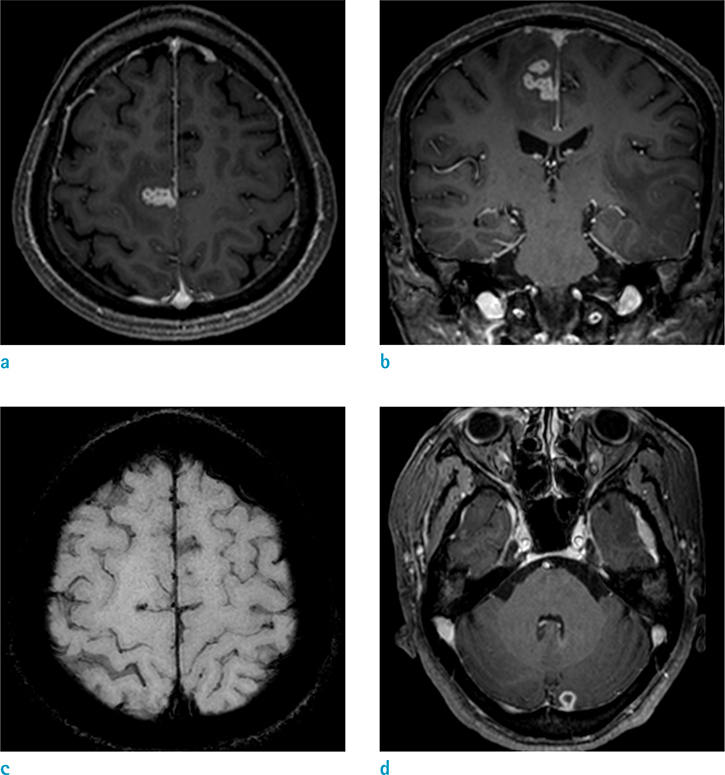
Fig. 2 Contrast-enhanced T1-weighted axial (a) and coronal (b) MRI showed multiple ring and nodular enhancing lesions in the cortico-sulcal area of the right medial frontoparietal lobe and left paramedian cerebellum (c). Some of these showed dark signal intensity on SWI (d).

Fig. 3 Follow-up MRI (4 weeks later) contrast-enhanced T1-weighted axial (a) and coronal (b) MRI, showed a marked decrease in the extent of the plaque-like enhancing mass of the left temporal subdural space (arrows). Also multiple tuberculomas in the right high frontoparietal cortico-sulcal area (c) and left paramedian cerebellum (d), showed a marked decrease in size and number on contrast-enhanced T1-weighted axial MRI.
Reference
-
1. Kwon YS, Koh WJ. Diagnosis of pulmonary tuberculosis and nontuberculous mycobacterial lung disease in Korea. Tuberc Respir Dis (Seoul). 2014; 77:1–5.2. Burrill J, Williams CJ, Bain G, Conder G, Hine AL, Misra RR. Tuberculosis: a radiologic review. Radiographics. 2007; 27:1255–1273.3. Khanna PC, Godinho S, Patkar DP, Pungavkar SA, Lawande MA. MR spectroscopy-aided differentiation: "giant" extra-axial tuberculoma masquerading as meningioma. AJNR Am J Neuroradiol. 2006; 27:1438–1440.4. Korea Centers for Disease Control and Prevention. Annual report on the notified tuberculosis in Korea. Cheongju (South Korea): The Centers;2015.5. Aggarwal A, Patra DP, Gupta K, Sodhi HB. Dural tuberculoma mimicking meningioma: a clinicoradiologic review of dural en-plaque lesions. World Neurosurg. 2016; 88:686.e1–686.e7.6. Srikanteswara PK, Pampapati PK, Yelsangikar KR. En-plaque central nervous system tuberculoma - an uncommon entity: clinico-radiological profile in a Cohort from a tertiary referral centre. J Clin Diagn Res. 2016; 10:OC11–OC14.7. Patkar D, Narang J, Yanamandala R, Lawande M, Shah GV. Central nervous system tuberculosis: pathophysiology and imaging findings. Neuroimaging Clin N Am. 2012; 22:677–705.8. Alkan A, Parlak M, Baysal T, Sigirci A, Kutlu R, Altinok T. Enplaque tuberculomas of tentorium in a pregnant woman: follow-up with MRI(2003:2b). Eur Radiol. 2003; 13:1190–1193.9. Lee JY. Diagnosis and treatment of extrapulmonary tuberculosis. Tuberc Respir Dis (Seoul). 2015; 78:47–45.10. Adachi K, Yoshida K, Tomita H, Niimi M, Kawase T. Tuberculoma mimicking falx meningioma--case report. Neurol Med Chir (Tokyo). 2004; 44:489–449.
