J Clin Neurol.
2016 Jul;12(3):371-372. 10.3988/jcn.2016.12.3.371.
Postictal Prosopometamorphopsia after Focal Status Epilepticus due to Cavernous Hemangioma in the Right Occipital Lobe
- Affiliations
-
- 1Department of Neurology, Seoul National University College of Medicine, Seoul National University Hospital, Seoul, Korea.
- 2Department of Neurology, Seoul National University College of Medicine, Seoul National University Bundang Hospital, Seongnam, Korea. nrpsh@snu.ac.kr
- KMID: 2354127
- DOI: http://doi.org/10.3988/jcn.2016.12.3.371
Abstract
- No abstract available.
Figure
-
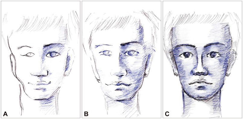
Fig. 1 Patient's drawing of his doctor's face. A: A drawing made on day 3 depicts bilateral prosopometamorphopsia (PM) that is more severe in the right half of the face. B: A drawing made on day 4 depicts unilateral PM restricted to the right half of the face. C: A drawing made on day 11 depicts a normal face.
Reference
-
1. Blom JD, Sommer IE, Koops S, Sacks OW. Prosopometamorphopsia and facial hallucinations. Lancet. 2014; 384:1998.
Article2. Heo K, Cho YJ, Lee SK, Park SA, Kim KS, Lee BI. Single-photon emission computed tomography in a patient with ictal metamorphopsia. Seizure. 2004; 13:250–253.
Article3. Dalrymple KA, Davies-Thompson J, Oruc I, Handy TC, Barton JJ, Duchaine B. Spontaneous perceptual facial distortions correlate with ventral occipitotemporal activity. Neuropsychologia. 2014; 59:179–191.
Article
- Full Text Links
-
- Actions
-
Cited
- CITED
-
- Close
- Share
-
- Similar articles
-
- Neuronal damage confirmed by 1H-MRS in occipital lobe complex partial status epilepticus
- 18F-FDG PET and 99mTc-ECD SPECT between Ictal and Interictal Phase in a Patient with Status Epilepticus Arising from the Occipital Lobe
- Cavernous Hemangioma on the Frontal Lobe
- Recurrent Cavernous Hemangioma of the Spermatic Cord
- Extensive Hemispheric Involvement on Diffusion-Weighted Image in a Patient with Status Epilepticus
