A Case of Primary Bilateral Angiosarcoma of the Breast
- Affiliations
-
- 1Department of Diagnostic Radiology, College of Medicine, Chosun University, Gwangju, Korea. yshkim@chosun.ac.kr
- 2Department of Pathology, College of Medicine, Chosun University, Gwangju, Korea.
- KMID: 2353681
- DOI: http://doi.org/10.3348/jksr.2016.75.4.300
Abstract
- Angiosarcoma is a malignant tumor of endovascular origin that can occur in any part of the body including the breast; however, angiosarcoma of the breast is quite rare. There are two sub-types of breast angiosarcoma. One of the subtypes, primary breast angiosarcoma, is very rare; only a few cases have been reported to date. Bilateral primary breast angiosarcoma is even rarer, and several cases have been reported in postmenopausal women. We report a case of a 34-year-old woman with primary bilateral angiosarcoma. She had no other significant medical history, such as breast surgery or radiotherapy, which can lead to secondary angiosarcoma of the breast.
MeSH Terms
Figure
-
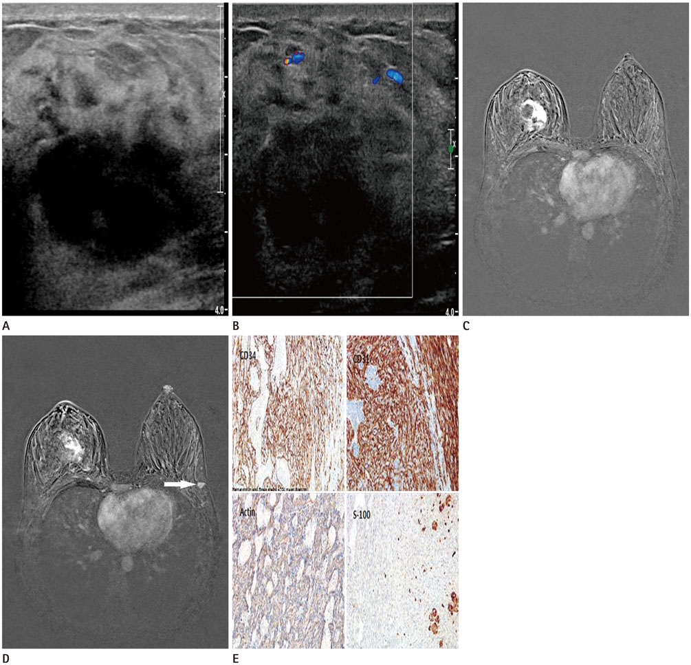
Fig. 1 A 34-year-old woman with primary bilateral angiosarcoma of the breast presenting with a palpable mass in the right breast. A. Ultrasonography shows a circumscribed hypoechoic mass at the 12 o'clock position in the right breast. B. On a color Doppler image, no increased vascularity is apparent around or within the mass. C. Axial contrast-enhanced T1 fat suppression magnetic resonance imaging (MRI) shows an irregular mass in the upper central portion of the right breast (60s after gadolinium injection). Multiple unenhanced portions are apparent in the mass. D. The lower level of the axial contrast-enhanced T1 fat suppression MRI scan shows a 9 mm enhancing oval shaped mass (arrow) in the lateral portion of the left breast. E. Tumor cells show immunoreactivity for CD34, CD31, and actin, but not for S-100 (400 × magnification).
Reference
-
1. Chen KT, Kirkegaard DD, Bocian JJ. Angiosarcoma of the breast. Cancer. 1980; 46:368–371.2. Kaklamanos IG, Birbas K, Syrigos KN, Vlachodimitropoulos D, Goutas N, Bonatsos G. Breast angiosarcoma that is not related to radiation exposure: a comprehensive review of the literature. Surg Today. 2011; 41:163–168.3. Kikawa Y, Konishi Y, Nakamoto Y, Harada T, Takeo M, Ogata M, et al. Angiosarcoma of the breast - specific findings of MRI. Breast Cancer. 2006; 13:369–373.4. Liberman L, Dershaw DD, Kaufman RJ, Rosen PP. Angiosarcoma of the breast. Radiology. 1992; 183:649–654.5. Pai MR, Upadhyaya K, Naik R, Malhotra S. Bilateral angiosarcoma breast diagnosed by fine needle aspiration cytology. Indian J Pathol Microbiol. 2008; 51:421–423.6. Sher T, Hennessy BT, Valero V, Broglio K, Woodward WA, Trent J, et al. Primary angiosarcomas of the breast. Cancer. 2007; 110:173–178.7. Rosen PP. Rosen's breast pathology. Philadelphia: Lippincott Williams & Wilkins;2001. p. 839.8. Sener SF, Milos S, Feldman JL, Martz CH, Winchester DJ, Dieterich M, et al. The spectrum of vascular lesions in the mammary skin, including angiosarcoma, after breast conservation treatment for breast cancer. J Am Coll Surg. 2001; 193:22–28.9. Schnarkowski P, Kessler M, Arnholdt H, Helmberger T. Angiosarcoma of the breast: mammographic, sonographic, and pathological findings. Eur J Radiol. 1997; 24:54–56.10. Yang WT, Hennessy BT, Dryden MJ, Valero V, Hunt KK, Krishnamurthy S. Mammary angiosarcomas: imaging findings in 24 patients. Radiology. 2007; 242:725–734.
