Well-Tolerated and Undiscovered Common Atrium until Late Adulthood
- Affiliations
-
- 1Division of Cardiology, Department of Internal Medicine, Anyang SAM Hospital, Anyang, Korea. fa5754@gmail.com
- KMID: 2353103
- DOI: http://doi.org/10.4250/jcu.2016.24.3.243
Abstract
- Common atrium is a rare congenital heart disease characterized by complete absence of the interatrial septum, and is commonly accompanied by malformation of the atrioventricular valve. Most patients with common atrium experience symptoms during childhood. Here, we describe a patient with common atrium who experienced his first obvious symptom at 48 years of age.
Keyword
Figure
-
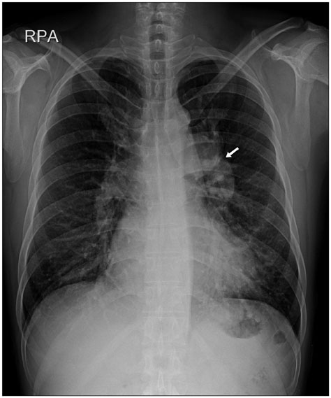
Fig. 1 Chest radiograph, showing cardiomegaly with a prominent left pulmonary trunk (arrow) and increased pulmonary vascularity.
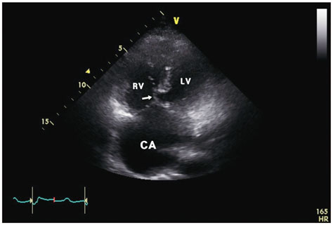
Fig. 2 Four-chamber transthoracic echocardiogram, showing complete absence of the interatrial septum. A chorda attached to the anterior common leaflet is seen (arrow). CA: common atrium, LV: left ventricle, RV: right ventricle.

Fig. 3 Four-chamber transesophageal echocardiogram, showing complete absence of the interatrial septum. CA: common atrium, LV: left ventricle, RV: right ventricle.
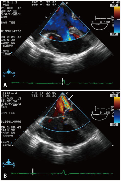
Fig. 4 Color Doppler transesophageal echocardiograms. A: Systolic phase. B: Tricuspid regurgitation between the right lateral leaflet and anterior common leaflet (arrowhead) is seen along with mild mitral regurgitation (arrow). No interventricular shunt is evident.
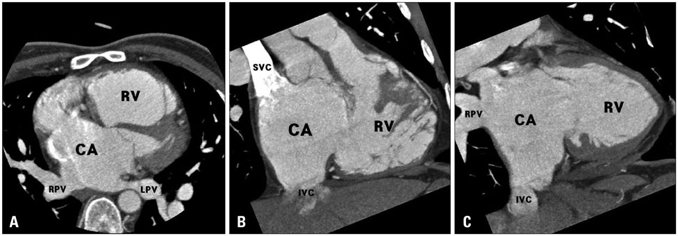
Fig. 5 Cardiac computed tomographic images. A: The right and left pulmonary veins drain to the left side of the common atrium. B and C: The superior vena cava and inferior vena cava normally drain to the right side of the common atrium. CA: common atrium, RV: right ventricle, RPV: right pulmonary vein, LPV: left pulmonary vein, SVC: superior vena cava, IVC: inferior vena cava.
Reference
-
1. Campbell M. Incidence of cardiac malformations at birth and later, and neonatal mortality. Br Heart J. 1973; 35:189–200.2. Muñoz-Armas S, Gorrín JR, Anselmi G, Hernández PB, Anselmi A. Single atrium. Embryologic, anatomic, electrocardiographic and other diagnostic features. Am J Cardiol. 1968; 21:639–652.3. Jiang H, Wang H, Wang Z, Zhu H, Zhang R. Surgical correction of common atrium without noncardiac congenital anomalies. J Card Surg. 2013; 28:580–586.4. Young AH, Robinson A. Some malformations of the human heart. Med Chron. 1907-1908; 47:96–106.5. Digilio MC, Marino B, Giannotti A, Dallapiccola B. Single atrium, atrioventricular canal/postaxial hexodactyly indicating Ellis-van Creveld syndrome. Hum Genet. 1995; 96:251–253.6. Ferdman DJ, Brady D, Rosenzweig EB. Common atrium and pulmonary vascular disease. Pediatr Cardiol. 2011; 32:595–598.7. Rastelli GC, Rahimtoola SH, Ongley PA, McGoon DC. Common atrium: anatomy, hemodynamics, and surgery. J Thorac Cardiovasc Surg. 1968; 55:834–841.8. Asuman Kaftan H, Tanriverdi H, Kuru O, Bir LS. An asymptomatic case with single atrium. Echocardiography. 2006; 23:701–703.9. Demirelli S, Fırtına S, Ermiş E, İnci S. Common atrium: a rare congenital heart anomaly. Turk Kardiyol Dern Ars. 2015; 43:579.10. Hasanin AM, Kinsara AJ. Single atrium associated with persistent left superior vena cava in asymptomatic adult: case report and review of literature. Congenit Heart Dis. 2008; 3:368–371.
- Full Text Links
-
- Actions
-
Cited
- CITED
-
- Close
- Share
-
- Similar articles
-
- What and How Do Psychotherapist Consider the Developmental Stages in Adult Psychotherapy?
- Methods and Causes of Completed Suicides According to Age and Gender
- Cross-Sectional and Longitudinal Examination of Insulin Sensitivity and Secretion across Puberty among Non-Hispanic Black and White Children
- Neuroleptic Malignant Syndrome Caused by Long Term Intake of Haloperidol
- A Case of Acquired Bilateral Nevus of Ota-like Macules Accompanying the Common Blue Nevus
