Botryoid Wilms' Tumor in a Child Presenting with Gross Hematuria: A Case Report
- Affiliations
-
- 1Department of Radiology and Research Institute of Radiological Science, Severance Children's Hospital, Yonsei University College of Medicine, Seoul, Korea. mjl1213@yuhs.ac
- 2Department of Pediatric Urology, Urological Science Institute, Severance Children's Hospital, Yonsei University College of Medicine, Seoul, Korea.
- KMID: 2349325
- DOI: http://doi.org/10.3348/jksr.2016.75.3.198
Abstract
- We report a unique case of botryoid Wilms' tumor with its characteristic imaging findings in a 5-month-old boy presenting with gross hematuria. In our case, ultrasonography revealed lobulated hyperechoic lesions filling the pelvicalyceal system without parenchymal invasion, mimicking a blood clot. However, magnetic resonance imaging (MRI) demonstrated the exact extent of the lesion with diffusion restriction and delayed enhancement suggestive of a tumor. Despite their rarity, botryoid Wilms' tumors should be included in the differential diagnosis of lobulated renal pelvic lesions presenting as gross hematuria in children, and MRI can suggest the diagnosis.
MeSH Terms
Figure
-
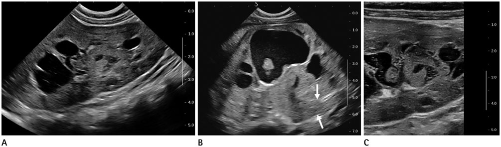
Fig. 1 Renal ultrasonography findings in a 5-month-old boy who presented with gross hematuria due to botryoid Wilms' tumor arising from the pelvis of the right kidney. A, B. Longitudinal views of the right kidney reveal (A) a lobulated hyperechoic mass filling the pelvicalyceal system without renal parenchymal invasion causing pelvicalyceal dilatation and (B) proximal ureteral extension (arrows) of the lesion. C. Using a high-frequency linear transducer, the pelvic mass is well delineated without evidence of renal parenchymal invasion.
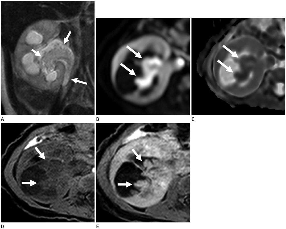
Fig. 2 Contrast enhanced MRI findings of botryoid Wilms' tumor. A. A coronal T2-weighted ultrafast spin echo sequence demonstrates a multilobulated soft tissue mass (arrows) with high signal intensity in the dilated right renal pelvis, lower pole calyx and proximal ureter, causing obstructive hydronephrosis of the right kidney. The right kidney also shows diffusely increased parenchymal signal intensity. B, C. The mass (arrows) demonstrates high signal intensity on the diffusion weighted image (B) and dark signal intensity on the apparent diffusion coefficient map (C), suggestive of diffusion restriction. D. An axial image of pre-contrast T1-weighted 3D fat-suppressed sequence demonstrates a mass (arrows) showing slightly high signal intensity. E. After gadolinium enhancement, the mass (arrows) shows delayed heterogeneous enhancement on a 5 minute delayed image of T1-weighted 3D fat-suppressed sequence.
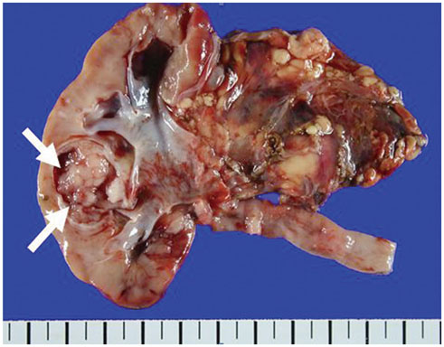
Fig. 3 The gross pathology of botryoid Wilm's tumor demonstrates a polypoid and whitish soft mass (arrows) in the renal calyx at the mid pole. The remaining renal parenchyma shows multifocal hemorrhage without gross tumor invasion.
Reference
-
1. Inoue M, Uchida K, Kohei O, Nashida Y, Deguchi T, Komada Y, et al. Teratoid Wilms' tumor: a case report with literature review. J Pediatr Surg. 2006; 41:1759–1763.2. Nagahara A, Kawagoe M, Matsumoto F, Tohda A, Shimada K, Yasui M, et al. Botryoid Wilms' tumor of the renal pelvis extending into the bladder. Urology. 2006; 67:845.e15–845.e17.3. Xu G, Hu J, Wu Y, Xiao Y, Xu M. Botryoid Wilms' tumor: a case report and review of the literature. World J Surg Oncol. 2013; 11:102.4. Honda A, Shima M, Onoe S, Hanada M, Nagai T, Nakajima S, et al. Botryoid Wilms tumor: case report and review of literature. Pediatr Nephrol. 2000; 14:59–61.5. Mizuno K, Hayashi Y, Tozawa K, Iwatsuki S, Kojima Y, Kohri K. Single-nucleotide polymorphism in WT1 gene in a hyperplastic intralobar nephrogenic rest with botryoid protrusion. Urology. 2010; 76:149–152.6. Tu BW, Ye WJ, Li YH. Botryoid Wilms' tumor: report of two cases. World J Pediatr. 2011; 7:274–276.7. Yanai T, Okazaki T, Yamataka A, Kobayashi H, Lane GJ, Saito M, et al. Botryoid Wilms' tumor: a report of two cases. Pediatr Surg Int. 2005; 21:43–46.8. Lowe LH, Isuani BH, Heller RM, Stein SM, Johnson JE, Navarro OM, et al. Pediatric renal masses: Wilms tumor and beyond. Radiographics. 2000; 20:1585–1603.9. Geller E, Kochan PS. Renal neoplasms of childhood. Radiol Clin North Am. 2011; 49:689–709, vi.10. Kim J. Ultrasonographic features of focal xanthogranulomatous pyelonephritis. J Ultrasound Med. 2004; 23:409–416.
