J Korean Ophthalmol Soc.
2016 Aug;57(8):1316-1319. 10.3341/jkos.2016.57.8.1316.
Use of Video-oculography in the Diagnosis of Superior Oblique Myokymia
- Affiliations
-
- 1Department of Ophthalmology, Chonbuk National University Medical School, Jeonju, Korea. ahnmin@jbnu.ac.kr
- 2Research Institute of Clinical Medicine, Chonbuk National University, Jeonju, Korea.
- 3Biomedical Research Institute, Chonbuk National University Hospital, Jeonju, Korea.
- KMID: 2349083
- DOI: http://doi.org/10.3341/jkos.2016.57.8.1316
Abstract
- PURPOSE
Superior oblique myokymia is intermittent spontaneous contractions of the superior oblique muscle presenting as rapid and small-amplitude intorsions and depressions of the eye. The authors report a case of superior oblique myokymia that was objectively and quantitatively diagnosed with slit lamp examination and video-oculography and completely resolved with medical treatment.
CASE SUMMARY
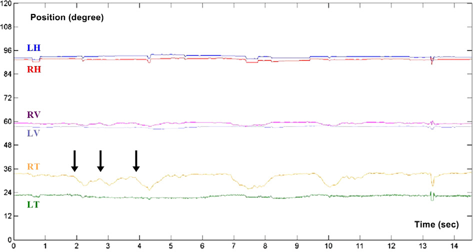
A 44-year-old woman presented with a seven-year history of intermittent oscillopsia which continued for few seconds. She had no history of head trauma or systemic ocular disease, and the anterior segment and fundus examination were unremarkable. Right eye intorsion lasting for a few seconds as detected by slit lamp examination. Eye movements were recorded using video-oculography, which showed a torsional nystagmus of 5 to 10 degrees with 2 to 5 vertical components in the right eye. Based on these findings, the patient was diagnosed with superior oblique myokymia. The patient was prescribed topical timolol ophthalmic solution, one drop twice per day, but the symptoms persisted. Timolol ophthalmic solution was stopped and replaced with carbamazepine, 200 mg twice a day, which resolved her symptoms.
CONCLUSIONS
Slit lamp examination and video-oculography can be used as objective and quantitative diagnostic tools in order to confirmed a diagnosis and lead to proper treatment.
MeSH Terms
Figure
Reference
-
References
1. Duane A. Unilateral rotary nystagmus. Trans Am Ophthalmol Soc. 1906; 11(Pt 1):63–7.2. Hoyt WF, Keane JR. Superior oblique myokymia. Report and abdominal on five cases of benign intermittent uniocular microtremor. Arch Ophthalmol. 1970; 84:461–7.3. Kim JS, Yun CH, Kim KS. A case of superior oblique myokymia. J Korean Neurol Assoc. 2002; 20:404–6.4. Lee SH, Kim DG, You JH, Song BR. A case of superior oblique myokymia. J Korean Ophthalmol Soc. 1997; 38:2053–5.5. Tomsak RL, Kosmorsky GS, Leigh RJ. Gabapentin attenuates abdominal oblique myokymia. Am J Ophthalmol. 2002; 133:721–3.6. Hashimoto M, Ohtsuka K, Suzuki Y, et al. Superior oblique myokymia caused by vascular compression. J Neuroophthalmol. 2004; 24:237–9.
Article7. Yousry I, Dieterich M, Naidich TP, et al. Superior oblique myokymia: magnetic resonance imaging support for the neuro-vascular compression hypothesis. Ann Neurol. 2002; 51:361–8.
Article8. Borgman CJ. Topical timolol in the treatment of monocular oscillopsia secondary to superior oblique myokymia: a review. J Optom. 2014; 7:68–74.
Article9. Williams PE, Purvin VA, Kawasaki A. Superior oblique myokymia: efficacy of medical treatment. J AAPOS. 2007; 11:254–7.
Article10. Ruttum MS, Harris GJ. Superior oblique myectomy and trochlear resection for superior oblique myokymia. Am J Ophthalmol. 2009; 148:563–5.
Article


