Evaluation of Clinical Applicability of Stress Radiography for Shoulder Instability: Correlation between the Degree of Displacement and the Physical Examinations
- Affiliations
-
- 1Global Center for Shoulder, Elbow & Sports, NEON Orthopaedic Clinic, Korea.
- 2Department of Orthopaedic Surgery, Konkuk University Medical Center, Seoul, Korea.
- 3Department of Orthopaedic Surgery, Inje University Haeundae Paik Hospital, Busan, Korea. neomed95@naver.com
- 4Institute of Statistics, Korea University, Seoul, Korea.
- KMID: 2348640
- DOI: http://doi.org/10.4055/jkoa.2016.51.4.327
Abstract
- PURPOSE
The purpose of this study is to examine the clinical applicability of stress radiography in patients presenting with shoulder instability.
MATERIALS AND METHODS
Fifty-six patients diagnosed with shoulder instability and 20 healthy volunteers participated in the study. Degree of displacement of the humeral head as measured on stress radiography using a Telos GA-IIE device was compared with the results of the physical examinations. Four types of stress radiography were captured while applying 15 daN of force anteriorly (AER0 and AER60) and posteriorly (PER0 and PER60) at two different positions: (1) 90° of abduction combined with 0° of external rotation, and (2) 90° of abduction combined with 60° of external rotation.
RESULTS
The degree of displacement of affected shoulders of 44 patients showed significantly larger displacement than normal shoulders (p<0.05), and the comparison between 56 affected shoulders of the patients and 40 normal shoulders of the volunteers showed significantly larger displacement only in PER0 and PER60 of the patients (p<0.05). Among the four radiographs of affected shoulders, AER60 showed significantly less displacement (p=0.046). The anterior drawer test under anesthesia of 16 patients who underwent surgery for anterior instability showed positive correlation with AER0 (Spearman's rho=0.56, p<0.024). Significantly larger anterior displacement of the load and shift test was observed in the subgroup with anterior displacement more than 3 mm (p=0.028), and higher positive frequency of the Kim's test was observed in the subgroup with posterior displacement more than 3 mm (p=0.005).
CONCLUSION
Stress radiography using a Telos GA-IIE device could discriminate the affected shoulder. Although it could not replace individual physical examinations, the degree of displacement correlates with some physical examinations for shoulder instability.
MeSH Terms
Figure
-

Figure 1 The Telos device mainly consists of a lower arm positioner for external rotation (1), shoulder fixation pads (2), and a pressure support (3).

Figure 2 Examples of anterior drawer stress radiography, AER0 and AER60. After seating the patient on the chair, the patient's arm is abducted by 90° and placed on the device, the axillary portion is placed in the middle of the cassette, and the coracoid process and scapular spine are fixed on the shoulder fixation pad. The pressure support pad is placed approximately 2 cm lateral to the margin of the acromion, and by using support devices, 15 daN pressure is steadily applied toward the back (abdomen), and under 60 Kvp and 10 mA film conditions, a beam is shot vertical to the acromioclavicular joint in the forward distal 30° direction with an source-image distance of 100 cm.
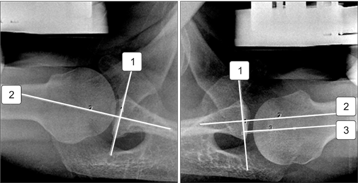
Figure 3 Example of posterior drawer stress radiography measurement in a patient. A line is drawn parallel to the glenoid articular surface (1), a line is drawn bisecting the glenoid articular surface perpendicular to line 1 (2), and a line is drawn from the rotation center of the humeral head perpendicular to line 1 (3). The distance between line 1 and 2 represents the posterior displacement.
Reference
-
1. Sein ML, Appleyard RC, Walton JR, Bradley T, Murrell GA. Reliability of a new shoulder laxometer to assess inferior glenohumeral joint translation. Br J Sports Med. 2008; 42:178–182.
Article2. Harryman DT 2nd, Sidles JA, Harris SL, Matsen FA 3rd. Laxity of the normal glenohumeral joint: a quantitative in vivo assessment. J Shoulder Elbow Surg. 1992; 1:66–76.
Article3. Borsa PA, Jacobson JA, Scibek JS, Dover GC. Comparison of dynamic sonography to stress radiography for assessing glenohumeral laxity in asymptomatic shoulders. Am J Sports Med. 2005; 33:734–741.
Article4. Papilion JA, Shall LM. Fluoroscopic evaluation for subtle shoulder instability. Am J Sports Med. 1992; 20:548–552.
Article5. Georgousis H, Ring D. Radiographic glenohumeral joint translation evaluated with a new shoulder positioning device. J Shoulder Elbow Surg. 1996; 5:S87.6. Ellenbecker TS, Mattalino AJ, Elam E, Caplinger R. Quantification of anterior translation of the humeral head in the throwing shoulder. Manual assessment versus stress radiography. Am J Sports Med. 2000; 28:161–167.7. Sauers EL, Borsa PA, Herling DE, Stanley RD. Instrumented measurement of glenohumeral joint laxity and its relationship to passive range of motion and generalized joint laxity. Am J Sports Med. 2001; 29:143–150.
Article8. Tzannes A, Paxinos A, Callanan M, Murrell GA. An assessment of the interexaminer reliability of tests for shoulder instability. J Shoulder Elbow Surg. 2004; 13:18–23.
Article9. Ellenbecker TS, Bailie DS, Mattalino AJ, et al. Intrarater and interrater reliability of a manual technique to assess anterior humeral head translation of the glenohumeral joint. J Shoulder Elbow Surg. 2002; 11:470–475.
Article10. Silliman JF, Hawkins RJ. Classification and physical diagnosis of instability of the shoulder. Clin Orthop Relat Res. 1993; 291:7–19.
Article11. Hawkins RJ, Bell RH. Shoulder instability: diagnosis and management. Can J Sport Sci. 1987; 12:67–70.12. Faber KJ, Homa K, Hawkins RJ. Translation of the glenohumeral joint in patients with anterior instability: awake examination versus examination with the patient under anesthesia. J Shoulder Elbow Surg. 1999; 8:320–323.13. Cofield RH, Irving JF. Evaluation and classification of shoulder instability. With special reference to examination under anesthesia. Clin Orthop Relat Res. 1987; 223:32–43.14. Gerber C, Ganz R. Clinical assessment of instability of the shoulder. With special reference to anterior and posterior drawer tests. J Bone Joint Surg Br. 1984; 66:551–556.
Article15. Hawkins RJ, Mohtadi NGH. Clinical evaluation of shoulder instability. Clin J Sport Med. 1991; 1:59–64.
Article16. Borsa PA, Sauers EL, Herling DE, Manzour WF. In vivo quantification of capsular end-point in the nonimpaired glenohumeral joint using an instrumented measurement system. J Orthop Sports Phys Ther. 2001; 31:419–426.17. Borsa PA, Scibek JS, Jacobson JA, Meister K. Sonographic stress measurement of glenohumeral joint laxity in collegiate swimmers and age-matched controls. Am J Sports Med. 2005; 33:1077–1084.
Article18. Tibone JE, Lee TQ, Csintalan RP, Dettling J, McMahon PJ. Quantitative assessment of glenohumeral translation. Clin Orthop Relat Res. 2002; 400:93–97.
Article19. Lippitt SB, Harris SL, Harryman DT 2nd, Sidles J, Matsen FA 3rd. In vivo quantification of the laxity of normal and unstable glenohumeral joints. J Shoulder Elbow Surg. 1994; 3:215–223.
Article20. Sauers EL, Borsa PA, Herling DE, Manzour WF, Stanley RD. Validity of an instrumented measurement technique for quantifying glenohumeral joint laxity and stiffness. J Athl Train. 2001; 36:S40.21. Sauers EL, Borsa PA, Herling DE, Stanley RD. Instrumented measurement of glenohumeral joint laxity: reliability and normative data. Knee Surg Sports Traumatol Arthrosc. 2001; 9:34–41.
Article22. Cain PR, Mutschler TA, Fu FH, Lee SK. Anterior stability of the glenohumeral joint. A dynamic model. Am J Sports Med. 1987; 15:144–148.23. Branch TP, Avilla O, London L, Hutton WC. Correlation of medial/lateral rotation of the humerus with glenohumeral translation. Br J Sports Med. 1999; 33:347–351.
Article24. O'Connell PW, Nuber GW, Mileski RA, Lautenschlager E. The contribution of the glenohumeral ligaments to anterior stability of the shoulder joint. Am J Sports Med. 1990; 18:579–584.25. Terry GC, Hammon D, France P, Norwood LA. The stabilizing function of passive shoulder restraints. Am J Sports Med. 1991; 19:26–34.
Article
- Full Text Links
-
- Actions
-
Cited
- CITED
-
- Close
- Share
-
- Similar articles
-
- Physical Examination of Shoulder Instability
- The Factors associated with Postural Control after Anterior Cruciate Ligament Reconstruction
- Correlation of the Rotator Interval on Direct CT Arthrography in Shoulder Instability Patients with External Rotation Stress Radiographs
- Instability Patterns of Normal Midcarpal and Radiocarpal Joint in the Sagittal Plane
- Physical Examinations of Rotator Cuff Tear
