Endoscopic Diagnosis and Differentiation of Inflammatory Bowel Disease
- Affiliations
-
- 1Department of Internal Medicine, St. Vincent's Hospital, College of Medicine, The Catholic University of Korea, Suwon, Korea. drmaloman@catholic.ac.kr
- KMID: 2348257
- DOI: http://doi.org/10.5946/ce.2016.090
Abstract
- Patients with inflammatory bowel disease have significantly increased in recent decades in Korea. Intestinal tuberculosis (ITB) and intestinal Behcet's disease (BD), which should be differentiated from Crohn's disease (CD), are more frequent in Korea than in the West. Thus, the accurate diagnosis of these inflammatory diseases is problematic in Korea and clinicians should fully understand their clinical and endoscopic characteristics. Ulcerative colitis mostly presents with rectal inflammation and continuous lesions, while CD presents with discontinuous inflammatory lesions and frequently involves the ileocecal area. Involvement of fewer than four segments, a patulous ileocecal valve, transverse ulcers, and scars or pseudopolyps are more frequently seen in ITB than in CD. A few ulcers with discrete margins are a typical endoscopic finding of intestinal BD. However, the differential diagnosis is difficult in many clinical situations because typical endoscopic findings are not always observed. Therefore, clinicians should also consider symptoms and laboratory, pathological, and radiological findings, in addition to endoscopic findings.
Keyword
MeSH Terms
Figure
-
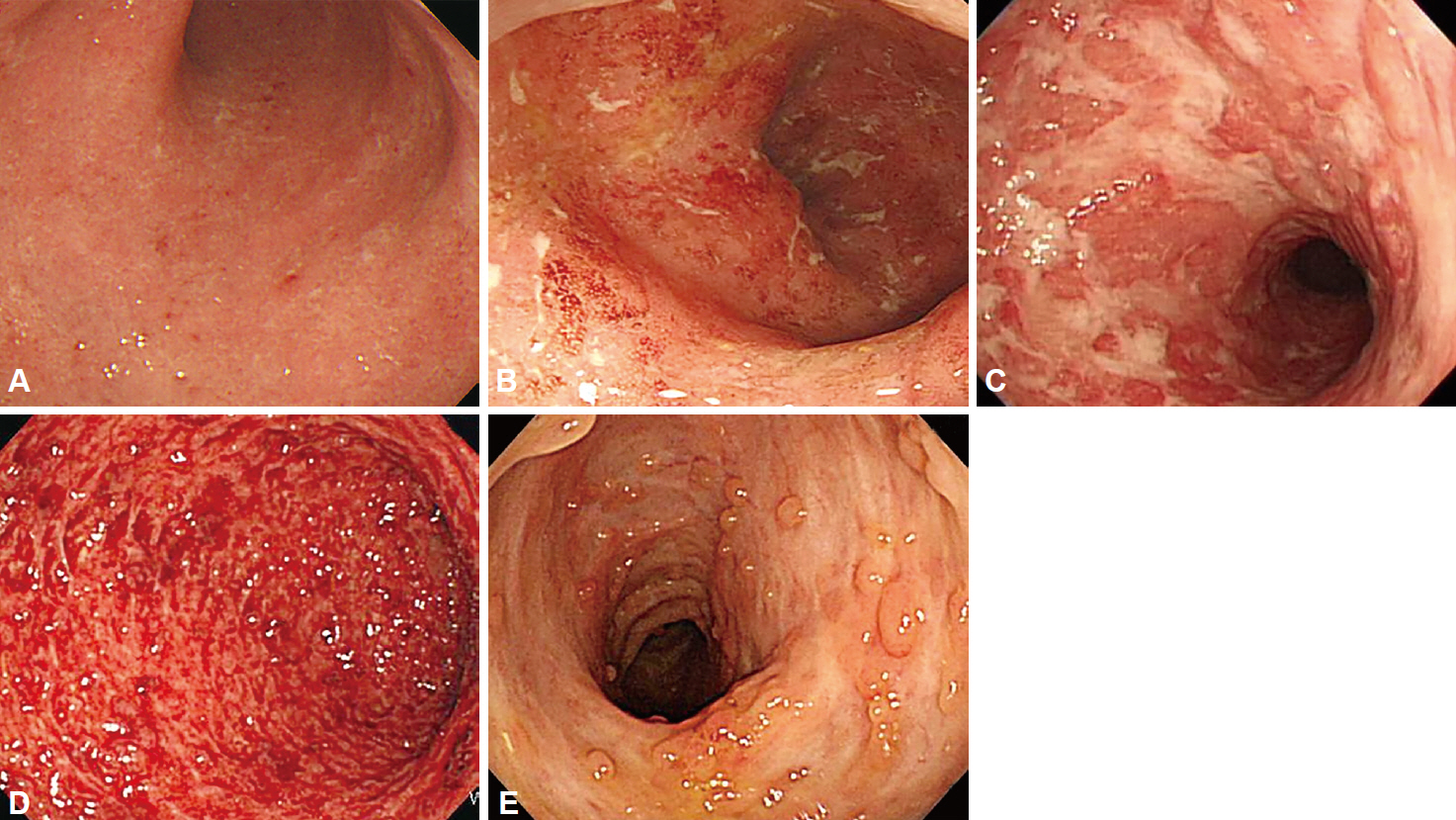
Fig. 1. Typical endoscopic features of ulcerative colitis. (A) Mild: mucosal erythema, fine granularity, decreased vascular marking. (B) Moderate: marked erythema, loss of vascular marking, erosions. (C) Severe: ulcers. (D) Severe: spontaneous bleeding. (E) Luminal narrowing with pseudopolyps.

Fig. 2. Typical endoscopic features of Crohn’s disease. (A) Longitudinal ulcers, (B) cobblestone appearance, (C) aphthous ulcers showing longitudinal array.
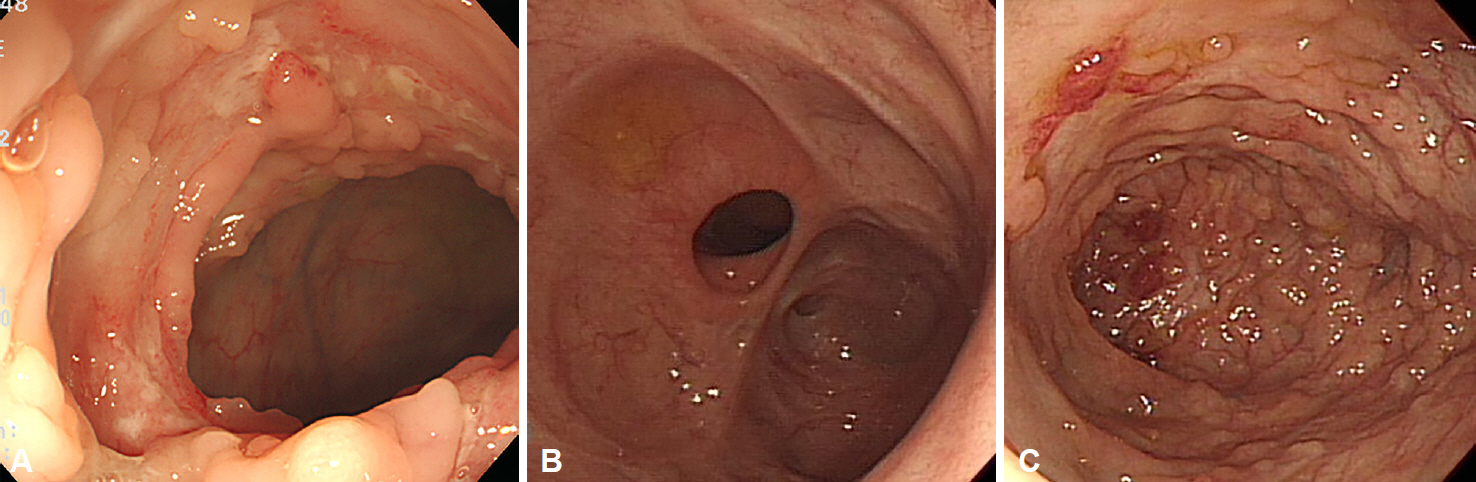
Fig. 3. Typical endoscopic features of intestinal tuberculosis. (A) Transverse ulcers, (B) deformed and patulous ileocecal valve, (C) pseudopolyps.
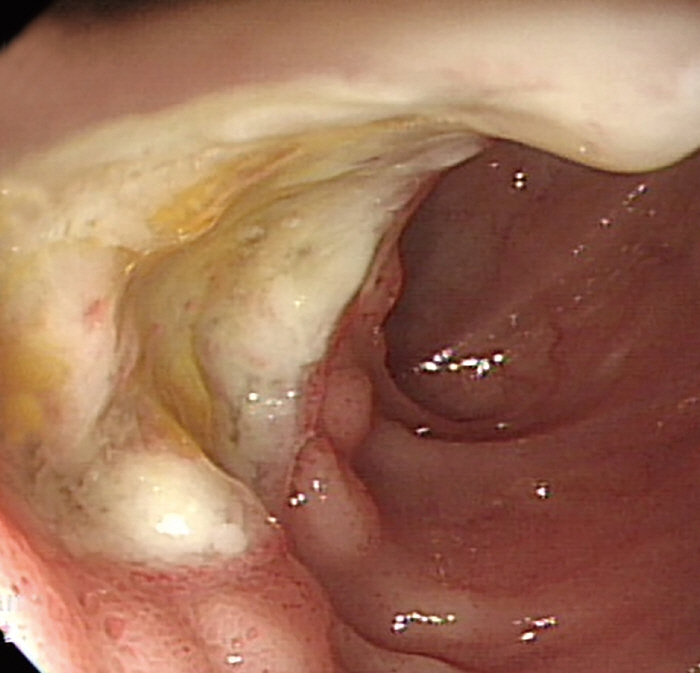
Fig. 4. Typical endoscopic feature of intestinal Behcet’s disease. A large, round, deep, and discrete ulcer in the terminal ileum.
Cited by 3 articles
-
Endoscopic Bamboo Joint-like Appearance of the Stomach in Crohn's Disease
Kwang Il Seo, Won Moon
Korean J Gastroenterol. 2017;69(2):151-154. doi: 10.4166/kjg.2017.69.2.151.Chronological Review of Endoscopic Indices in Inflammatory Bowel Disease
Joon Seop Lee, Eun Soo Kim, Won Moon
Clin Endosc. 2019;52(2):129-136. doi: 10.5946/ce.2018.042.Management of Crohn's disease in Taiwan: consensus guideline of the Taiwan Society of Inflammatory Bowel Disease
Shu-Chen Wei, Ting-An Chang, Te-Hsin Chao, Jinn-Shiun Chen, Jen-Wei Chou, Yenn-Hwei Chou, Chiao-Hsiung Chuang, Wen-Hung Hsu, Tien-Yu Huang, Tzu-Chi Hsu, Chun-Chi Lin, Hung-Hsin Lin, Jen-Kou Lin, Wei-Chen Lin, Yen-Hsuan Ni, Ming-Jium Shieh, I-Lun Shih, Chia-Tung Shun, Yuk-Ming Tsang, Cheng-Yi Wang, Horng-Yuan Wang, Meng-Tzu Weng, Deng-Chyang Wu, Wen-Chieh Wu, Hsu-Heng Yen, Jau-Min Wong
Intest Res. 2017;15(3):285-310. doi: 10.5217/ir.2017.15.3.285.
Reference
-
1. Yang SK, Yun S, Kim JH, et al. Epidemiology of inflammatory bowel disease in the Songpa-Kangdong district, Seoul, Korea, 1986-2005: a KASID study. Inflamm Bowel Dis. 2008; 14:542–549.
Article2. Suzuki Kurokawa M, Suzuki N. Behcet’s disease. Clin Exp Med. 2004; 4:10–20.
Article3. Kim YS, Kim YH, Lee KM, Kim JS, Park YS; IBD Study Group of the Korean Association of the Study of Intestinal Diseases. Diagnostic guideline of intestinal tuberculosis. Korean J Gastroenterol. 2009; 53:177–186.4. Choi CH, Jung SA, Lee BI, et al. Diagnostic guideline of ulcerative colitis. Korean J Gastroenterol. 2009; 53:145–160.5. Ye BD, Jang BI, Jeen YT, et al. Diagnostic guideline of Crohn’s disease. Korean J Gastroenterol. 2009; 53:161–176.6. Cheon JH, Shin SJ, Kim SW, et al. Diagnosis of intestinal Behcet’s disease. Korean J Gastroenterol. 2009; 53:187–193.7. Waye JD. The role of colonoscopy in the differential diagnosis of inflammatory bowel disease. Gastrointest Endosc. 1977; 23:150–154.
Article8. Waye JD. Endoscopy in inflammatory bowel disease: indications and differential diagnosis. Med Clin North Am. 1990; 74:51–65.
Article9. Jalan KN, Walker RJ, Sircus W, McManus JP, Prescott RJ, Card WI. Pseudopolyposis in ulcerative colitis. Lancet. 1969; 2:555–559.
Article10. D’Haens G, Geboes K, Peeters M, Baert F, Ectors N, Rutgeerts P. Patchy cecal inflammation associated with distal ulcerative colitis: a prospective endoscopic study. Am J Gastroenterol. 1997; 92:1275–1279.11. Park SH, Yang SK, Park SK, et al. Atypical distribution of inflammation in newly diagnosed ulcerative colitis is not rare. Can J Gastroenterol Hepatol. 2014; 28:125–130.
Article12. Haskell H, Andrews CW Jr, Reddy SI, et al. Pathologic features and clinical significance of “backwash” ileitis in ulcerative colitis. Am J Surg Pathol. 2005; 29:1472–1481.
Article13. Stange EF, Travis SP, Vermeire S, et al. European evidence based consensus on the diagnosis and management of Crohn’s disease: definitions and diagnosis. Gut. 2006; 55 Suppl 1:i1–i15.
Article14. Yao T. New criteria for the diagnosis of Crohn’s disease. Stomach Intest. 1996; 31:451–464.15. Park JB, Yang SK, Myung SJ, et al. Clinical characteristics at diagnosis and course of Korean patients with Crohn’s disease. Korean J Gastroenterol. 2004; 43:8–17.16. Kang MS, Park DI, Park JH, et al. Bamboo joint-like appearance of stomach in Korean patients with Crohn’s disease. Korean J Gastroenterol. 2006; 48:395–400.17. Yokota K, Saito Y, Einami K, et al. A bamboo joint-like appearance of the gastric body and cardia: possible association with Crohn’s disease. Gastrointest Endosc. 1997; 46:268–272.
Article18. Naga MI, Okasha HH, Ismail Z, El-Fatatry M, Hassan S, Monir BE. Endoscopic diagnosis of colonic tuberculosis. Gastrointest Endosc. 2001; 53:789–793.
Article19. Singh V, Kumar P, Kamal J, Prakash V, Vaiphei K, Singh K. Clinicocolonoscopic profile of colonic tuberculosis. Am J Gastroenterol. 1996; 91:565–568.20. Hoshino M, Shibata M, Goto N, et al. A clinical study of tuberculous colitis. Gastroenterol Jpn. 1979; 14:299–305.
Article21. Lee YJ, Yang SK, Myung SJ, et al. The usefulness of colonoscopic biopsy in the diagnosis of intestinal tuberculosis and pattern of concomitant extra-intestinal tuberculosis. Korean J Gastroenterol. 2004; 44:153–159.22. Podolsky DK. Inflammatory bowel disease. N Engl J Med. 2002; 347:417–429.
Article23. Lee HS, Choe J, Lee HJ, et al. Change in the diagnosis of inflammatory bowel disease: a hospital-based cohort study from Korea. Intest Res. 2016; 14:258–263.
Article24. Kim ES, Chen M, Lee J, Lee CK, Kim YS. Diagnosis of inflammatory bowel disease in Asia: the results of a multinational web-based survey in the 2nd Asian Organization for Crohn’s and Colitis (AOCC) meeting in Seoul. Intest Res. 2016; 14:224–230.25. Reese GE, Constantinides VA, Simillis C, et al. Diagnostic precision of anti-Saccharomyces cerevisiae antibodies and perinuclear antineutrophil cytoplasmic antibodies in inflammatory bowel disease. Am J Gastroenterol. 2006; 101:2410–2422.
Article26. Nikolaus S, Schreiber S. Diagnostics of inflammatory bowel disease. Gastroenterology. 2007; 133:1670–1689.
Article27. Lee YJ, Yang SK, Byeon JS, et al. Analysis of colonoscopic findings in the differential diagnosis between intestinal tuberculosis and Crohn’s disease. Endoscopy. 2006; 38:592–597.
Article28. Alvares JF, Devarbhavi H, Makhija P, Rao S, Kottoor R. Clinical, colonoscopic, and histological profile of colonic tuberculosis in a tertiary hospital. Endoscopy. 2005; 37:351–356.
Article29. Kim KM, Lee A, Choi KY, Lee KY, Kwak JJ. Intestinal tuberculosis: clinicopathologic analysis and diagnosis by endoscopic biopsy. Am J Gastroenterol. 1998; 93:606–609.
Article30. Kim YS, Kim YH, Kim WH, et al. Diagnostic utility of anti-Saccharomyces cerevisiae antibody (ASCA) and interferon-γ assay in the differential diagnosis of Crohn’s disease and intestinal tuberculosis. Clin Chim Acta. 2011; 412:1527–1532.
Article31. Lee SK, Kim BK, Kim TI, Kim WH. Differential diagnosis of intestinal Behcet’s disease and Crohn’s disease by colonoscopic findings. Endoscopy. 2009; 41:9–16.32. Cheon JH, Kim ES, Shin SJ, et al. Development and validation of novel diagnostic criteria for intestinal Behçet’s disease in Korean patients with ileocolonic ulcers. Am J Gastroenterol. 2009; 104:2492–2499.
Article
- Full Text Links
-
- Actions
-
Cited
- CITED
-
- Close
- Share
-
- Similar articles
-
- Diagnostic Tips for Making the Diagnosis of Inflammatory Bowel Disease
- Small Bowel Endoscopy in Inflammatory Bowel Disease
- Role of Advanced Endoscopic Imaging Techniques in the Management of Inflammatory Bowel Disease
- Differential Diagnosis of Inflammatory Bowel Disease: What Is the Role of Colonoscopy?
- Chronological Review of Endoscopic Indices in Inflammatory Bowel Disease
