Comparison of the Thickness of the Lamina Cribrosa and Vascular Factors in Early Normal-tension Glaucoma with Low and High Intraocular Pressures
- Affiliations
-
- 1Cheil Eye Hospital and Cheil Eye Research Institute, Daegu, Korea. 10041419@naver.com
- KMID: 2345824
- DOI: http://doi.org/10.3341/kjo.2014.28.6.473
Abstract
- PURPOSE
To compare the thickness of the lamina cribrosa (LC) and vascular factors of early normal-tension glaucoma (NTG) patients with high and low intraocular pressure (IOP) that are expected to be associated with the development of glaucoma.
METHODS
Seventy-one Korean NTG patients with low IOP (the highest IOP <15 mmHg, 40 patients) and high IOP (the lowest IOP >15 mmHg, 31 patients) were included in this study. The thickness of LC and vascular factors were compared. The thickness of the LC was measured using the enhanced depth imaging method with spectral domain optical coherence tomography (Heidelberg Spectralis).
RESULTS
The mean thickness of the central LC was 190.0 +/- 19.2 microm in the low IOP group and 197.8 +/- 23.6 microm in the high IOP group, but there was no statistical significant difference between the two groups (p > 0.05). The prevalence of self-reported Raynaud phenomenon was significantly higher in the low IOP group (33.0%) than the high IOP group (10.3%, p = 0.04).
CONCLUSIONS
The laminar thickness did not significantly differ between the high and low IOP groups. However, the prevalence of Raynaud phenomenon was higher in the low IOP groups. These results suggest that the development of glaucoma with low IOP patients may be more influenced by peripheral vasospasm, such as Raynaud phenomenon, rather than laminar thickness in NTG.
Keyword
MeSH Terms
-
Aged
Cross-Sectional Studies
Female
Humans
*Intraocular Pressure
Low Tension Glaucoma/*diagnosis
Male
Middle Aged
Nerve Fibers/pathology
Optic Disk/*pathology
Optic Nerve Diseases/*diagnosis
Raynaud Disease/*diagnosis
Retinal Ganglion Cells/pathology
Tomography, Optical Coherence
Tonometry, Ocular
Vision Disorders/diagnosis
Visual Fields
Figure
-
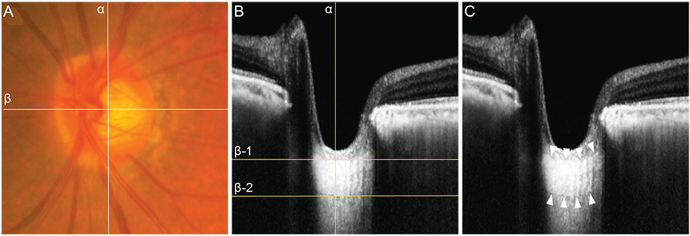
Fig. 1 (A) Fundus photograph and (B,C) spectral-domain optical coherence tomography (OCT) images in the enhanced depth imaging (EDI) mode in a 78-year-old woman with normal-tension glaucoma. The mean intraocular pressure was 10 mmHg. In (A), the center of the optic nerve head (ONH) was identified at the point where the vertical center (line α) of the ONH met the horizontal center from which the trunk of the central retinal vessels came out from the ONH (line β). In (B), in the horizontal cross-sectional view of the EDI-OCT, the lamina cribrosa thickness (204 µm) was measured along line α, between lines β-1 and β-2. In (C), both the anterior and posterior borders (delineated with white arrows) of the lamina cribrosa were identified in the EDI-OCT.
Reference
-
1. Cartwright MJ, Anderson DR. Correlation of asymmetric damage with asymmetric intraocular pressure in normal-tension glaucoma (low-tension glaucoma). Arch Ophthalmol. 1988; 106:898–900.2. Crichton A, Drance SM, Douglas GR, Schulzer M. Unequal intraocular pressure and its relation to asymmetric visual field defects in low-tension glaucoma. Ophthalmology. 1989; 96:1312–1314.3. Haefliger IO, Hitchings RA. Relationship between asymmetry of visual field defects and intraocular pressure difference in an untreated normal (low) tension glaucoma population. Acta Ophthalmol (Copenh). 1990; 68:564–567.4. Jonas JB, Grundler AE, Gonzales-Cortes J. Pressure-dependent neuroretinal rim loss in normal-pressure glaucoma. Am J Ophthalmol. 1998; 125:137–144.5. Wang XH, Stewart WC, Jackson GJ. Differences in optic discs in low-tension glaucoma patients with relatively low or high pressures. Acta Ophthalmol Scand. 1996; 74:364–367.6. Kim DM, Seo JH, Kim SH, Hwang SS. Comparison of localized retinal nerve fiber layer defects between a low-teen intraocular pressure group and a high-teen intraocular pressure group in normal-tension glaucoma patients. J Glaucoma. 2007; 16:293–296.7. Bellezza AJ, Rintalan CJ, Thompson HW, et al. Deformation of the lamina cribrosa and anterior scleral canal wall in early experimental glaucoma. Invest Ophthalmol Vis Sci. 2003; 44:623–637.8. Burgoyne CF, Downs JC. Premise and prediction-how optic nerve head biomechanics underlies the susceptibility and clinical behavior of the aged optic nerve head. J Glaucoma. 2008; 17:318–328.9. Inoue R, Hangai M, Kotera Y, et al. Three-dimensional high-speed optical coherence tomography imaging of lamina cribrosa in glaucoma. Ophthalmology. 2009; 116:214–222.10. Strouthidis NG, Grimm J, Williams GA, et al. A comparison of optic nerve head morphology viewed by spectral domain optical coherence tomography and by serial histology. Invest Ophthalmol Vis Sci. 2010; 51:1464–1474.11. Lee EJ, Kim TW, Weinreb RN, et al. Visualization of the lamina cribrosa using enhanced depth imaging spectral-domain optical coherence tomography. Am J Ophthalmol. 2011; 152:87–95.e1.12. Drance SM, Sweeney VP, Morgan RW, Feldman F. Studies of factors involved in the production of low tension glaucoma. Arch Ophthalmol. 1973; 89:457–465.13. Rojanapongpun P, Drance SM. The response of blood flow velocity in the ophthalmic artery and blood flow of the finger to warm and cold stimuli in glaucomatous patients. Graefes Arch Clin Exp Ophthalmol. 1993; 231:375–377.14. Gasser P. Ocular vasospasm: a risk factor in the pathogenesis of low-tension glaucoma. Int Ophthalmol. 1989; 13:281–290.15. Shirakashi M, Funaki S, Funaki H, et al. Measurement of retinal nerve fibre layer by scanning laser polarimetry and high pass resolution perimetry in normal tension glaucoma with relatively high or low intraocular pressure. Br J Ophthalmol. 1999; 83:353–357.16. Fleiss JL. The design and analysis of clinical experiments. New York: Wiley;1986. p. 1–32.17. Anderson DR, Hendrickson A. Effect of intraocular pressure on rapid axoplasmic transport in monkey optic nerve. Invest Ophthalmol. 1974; 13:771–783.18. Quigley H, Anderson DR. The dynamics and location of axonal transport blockade by acute intraocular pressure elevation in primate optic nerve. Invest Ophthalmol. 1976; 15:606–616.19. Downs JC, Roberts MD, Burgoyne CF. Mechanical environment of the optic nerve head in glaucoma. Optom Vis Sci. 2008; 85:425–435.20. Sigal IA, Flanagan JG, Tertinegg I, Ethier CR. Predicted extension, compression and shearing of optic nerve head tissues. Exp Eye Res. 2007; 85:312–322.21. Sigal IA, Flanagan JG, Tertinegg I, Ethier CR. Modeling individual-specific human optic nerve head biomechanics. Part I: IOP-induced deformations and influence of geometry. Biomech Model Mechanobiol. 2009; 8:85–98.22. Park SC, De Moraes CG, Teng CC, et al. Enhanced depth imaging optical coherence tomography of deep optic nerve complex structures in glaucoma. Ophthalmology. 2012; 119:3–9.23. Park HY, Jeon SH, Park CK. Enhanced depth imaging detects lamina cribrosa thickness differences in normal tension glaucoma and primary open-angle glaucoma. Ophthalmology. 2012; 119:10–20.24. Kondo Y, Niwa Y, Yamamoto T, et al. Retrobulbar hemodynamics in normal-tension glaucoma with asymmetric visual field change and asymmetric ocular perfusion pressure. Am J Ophthalmol. 2000; 130:454–460.25. Park HY, Jung KI, Na KS, et al. Visual field characteristics in normal-tension glaucoma patients with autonomic dysfunction and abnormal peripheral microcirculation. Am J Ophthalmol. 2012; 154:466–475.26. Tielsch JM, Katz J, Sommer A, et al. Hypertension, perfusion pressure, and primary open-angle glaucoma: a population-based assessment. Arch Ophthalmol. 1995; 113:216–221.27. Flammer J, Mozaffarieh M. What is the present pathogenetic concept of glaucomatous optic neuropathy? Surv Ophthalmol. 2007; 52:Suppl 2. S162–S173.28. Karakucuk S, Goktas S, Aksu M, et al. Ocular blood flow in patients with obstructive sleep apnea syndrome (OSAS). Graefes Arch Clin Exp Ophthalmol. 2008; 246:129–134.
- Full Text Links
-
- Actions
-
Cited
- CITED
-
- Close
- Share
-
- Similar articles
-
- Lamina Cribrosa Thickness in the Fellow Eyes of Patients with Unilateral Retinal Vein Occlusion
- Inter-eye Comparison of the Lamina Cribrosa Depth in Patients with Bilateral Normal-tension Glaucoma with Asymmetrical Damage
- Correlation between Trans-lamina Cribrosa Pressure Difference and Morphologic Parameters of Optic Disc in Normal Tension Glaucoma Patients
- Reversal of Optic Disc Cupping in Adults with Advanced Glaucoma
- Analysis of Systemic Risk Factorsin Normal Tension Glaucoma
