Gastric Mixed Adenoneuroendocrine Carcinoma with Squamous Differentiation: A Case Report
- Affiliations
-
- 1Department of Pathology, Kyungpook National University Hospital, Kyungpook National University Medical Center, Kyungpook National University School of Medicine, Daegu, Korea. san_0729@naver.com
- 2Department of Surgery, Gastric Cancer Center, Kyungpook National University Medical Center, Kyungpook National University School of Medicine, Daegu, Korea.
- 3Department of Pathology, Samsung Changwon Hospital, Sungkyunkwan University School of Medicine, Changwon, Korea.
- KMID: 2345558
- DOI: http://doi.org/10.4132/jptm.2015.10.17
Abstract
- No abstract available.
Figure
-
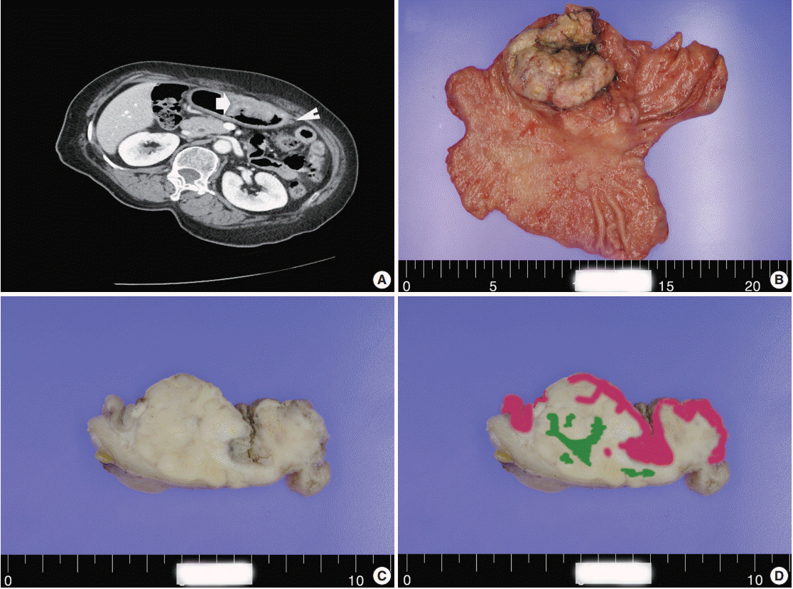
Fig. 1. (A) A computed tomography scan demonstrating a large ulcerofungating hypodense mass in the anterior gastric wall (arrow) and enlargement of multiple perigastric lymph nodes (arrowhead). Macroscopic findings (B) and cross-section of gastric cancer (C). (D) Mapping of the gastric mixed adenoneuroendocrine carcinoma with trilineage histologic differentiation composed of adenocarcinoma (pink color), large cell neuroendocrine carcinoma, and squamous cell carcinoma (green color).
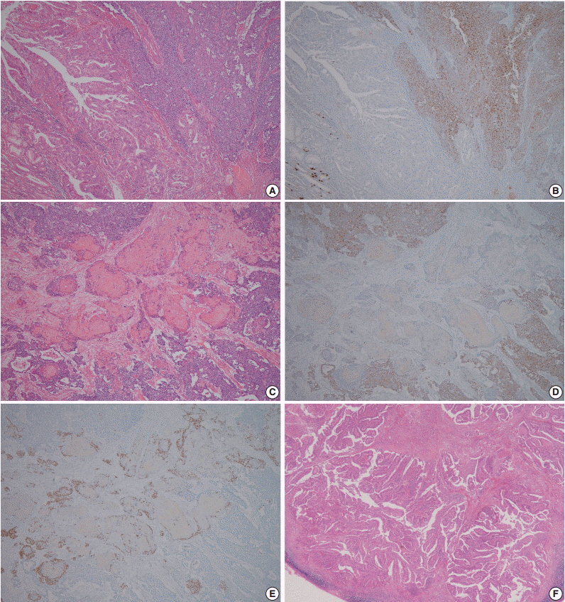
Fig. 2. Representative microscopic findings of mixed adenoneuroendocrine carcinoma with trilineage histologic differentiation. (A) Moderately differentiated adenocarcinoma admixed with large cell neuroendocrine carcinoma (NEC). (B) The NEC component demonstrates positive staining for chromogranin A, whereas the adenocarcinoma component shows negativity. (C) Squamous cell carcinoma (SqCC) component admixed with the NEC component, and (D) the NEC component presented positivity for chromogranin A, but not the SqCC component. (E) The cells with squamous cell differentiation are identified by expression of p63. (F) Metastasis of only the adenocarcinoma component to the gastric regional lymph nodes.

Fig. 3. Photomicrographs of immunohistochemistry (IHC) for c-erbB2 in mixed adenoneuroendocrine carcinoma with trilineage histologic differentiation. IHC for c-erbB2 shows strong positivity in the adenocarcinoma component (A), and in the metastatic regional lymph nodes (B), whereas IHC for c-erbB2 is negative in neuroendocrine carcinoma (C) and squamous cell carcinoma component (D).
Cited by 1 articles
-
Multiregion Comprehensive Genomic Profiling of a Gastric Mixed Neuroendocrine-Nonneuroendocrine Neoplasm with Trilineage Differentiation
Faheem Farooq, Kevin Zarrabi, Keith Sweeney, Joseph Kim, Jela Bandovic, Chiraag Patel, Minsig Choi
J Gastric Cancer. 2018;18(2):200-207. doi: 10.5230/jgc.2018.18.e16.
Reference
-
1. Bosman FT, Carneiro F, Hruban RH, Theise ND. WHO classification of tumours of the digestive system. 4th ed. Lyon: IARC Press;2010.2. Volante M, Rindi G, Papotti M. The grey zone between pure (neuro)endocrine and non-(neuro)endocrine tumours: a comment on concepts and classification of mixed exocrine-endocrine neoplasms. Virchows Arch. 2006; 449:499–506.
Article3. Haratake J, Horie A, Inoshita S. Gastric small cell carcinoma with squamous and neuroendocrine differentiation. Pathology. 1992; 24:116–20.
Article4. Shibuya H, Azumi N, Abe F. Gastric small-cell undifferentiated carcinoma with adeno and squamous cell carcinoma components. Acta Pathol Jpn. 1985; 35:473–80.5. Pericleous M, Toumpanakis C, Lumgair H, et al. Gastric mixed adenoneuroendocrine carcinoma with a trilineage cell differentiation: case report and review of the literature. Case Rep Oncol. 2012; 5:313–9.
Article6. Bartley AN, Rashid A, Fournier KF, Abraham SC. Neuroendocrine and mucinous differentiation in signet ring cell carcinoma of the stomach: evidence for a common cell of origin in composite tumors. Hum Pathol. 2011; 42:1420–9.
Article7. Kim KM, Kim MJ, Cho BK, Choi SW, Rhyu MG. Genetic evidence for the multi-step progression of mixed glandular-neuroendocrine gastric carcinomas. Virchows Arch. 2002; 440:85–93.
Article
- Full Text Links
-
- Actions
-
Cited
- CITED
-
- Close
- Share
-
- Similar articles
-
- Mixed adenoneuroendocrine carcinoma of the ampulla of Vater: Three case reports and a literature review
- Mixed Adenoneuroendocrine Gastric Carcinoma: A Case Report and Review of the Literature
- Multiregion Comprehensive Genomic Profiling of a Gastric Mixed Neuroendocrine-Nonneuroendocrine Neoplasm with Trilineage Differentiation
- Three Cases of Primary Adenosquamous Carcinoma of Stomach
- Mixed adenoneuroendocrine carcinoma in the stomach: a case report with a literature review
