MR Imaging of Lymphoepithelial Carcinoma in the Posterior Cervical Space: A Case Report
- Affiliations
-
- 1Department of Radiology, Eulji General Hospital, Eulji University School of Medicine, Seoul, Korea. kwchung99@eulji.ac.kr
- 2Department of Anatomic Pathology, Eulji General Hospital, Eulji University School of Medicine, Seoul, Korea.
Abstract
- Lymphoepithelial carcinoma is a rare category of malignant neoplasm that consists of undifferentiated malignant epithelial cells admixed with lymphocytes. Lymphoepithelial carcinoma predominantly occurs in the upper aero-digestive tract and only few cases of metastasis in the posterior cervical space have currently been reported. We report here on a case of lymphoepithelial carcinoma in the posterior cervical space.
Figure
-
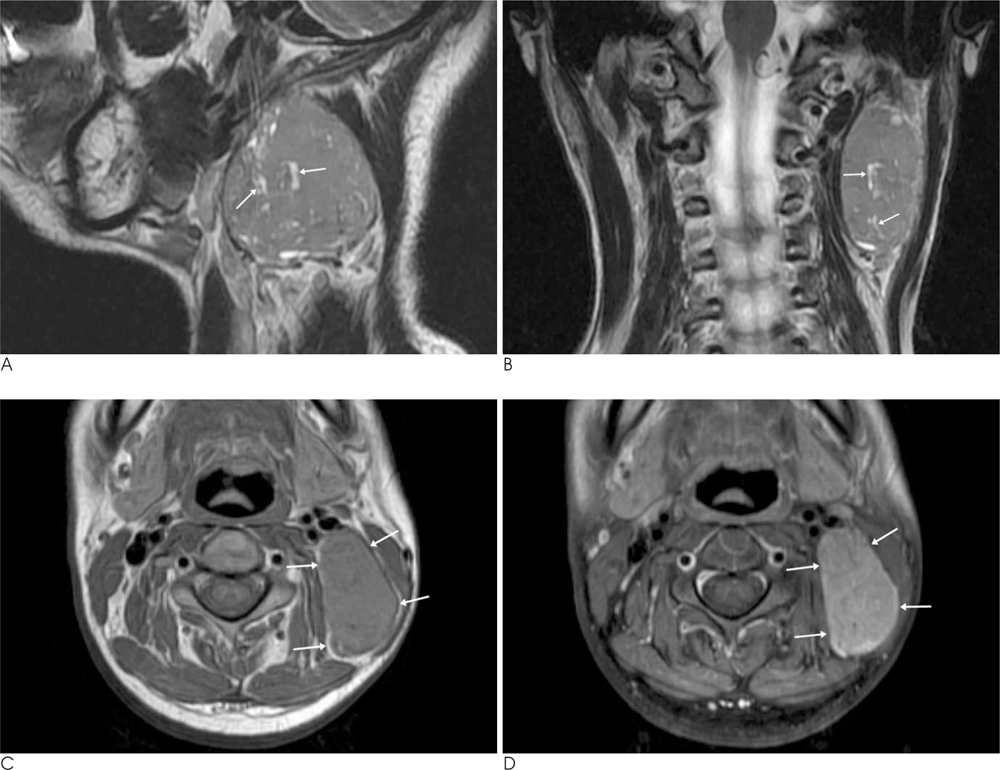
Fig. 1 The MR imaging reveals a well-defined mass with a lobulated contour in the left posterior cervical space. A, B. On the coronal (A) and sagittal (B) T2-weighted fast spin-echo MR images, the mass shows slightly higher signal intensity than the surrounding muscle and intra-tumoral microtubular cystic clefts (arrows). C. The axial T1-weighted spin-echo MR image shows an isointense mass (arrows) relative to the surrounding muscle. D. The gadolinium-enhanced axial fat suppressed T1-weighted spin-echo MR image shows the slightly heterogeneous enhancement of the mass (arrows) without central necrosis or an enhancing capsule.
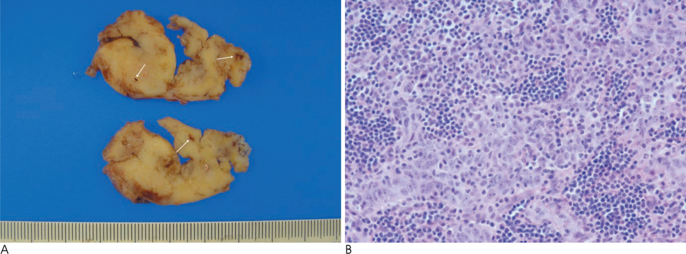
Fig. 2 The gross photography of the specimen (A) shows multiple dilated lymphovascular channels (arrows) in the tumor. The microscopic examination (B) shows islands of undifferentiated carcinoma cells admixed with many small lymphocytes and plasma cells (H & E staining, ×400).
Reference
-
1. Mills SE, Fechner RE. The nose, paranasal sinuses and nasopharynx. Diagnostic surgical pathology. 3nd ed. New York: Lippincott Williams & Wilkins;1999. p. 885–924.2. Chan JKC, Bray F, McCarron P, Foo W, Lee AWM, Yip T, et al. Tumours of the nasopharynx: Nasopharyngeal carcinoma. In : Bames L, Eveson JW, Reichart P, Sidransky D, editors. Pathology and genetics of head and neck tumours: WHO classification of tumours. Lyon: IARC Press;2005. p. 83–97.3. Wassef M, Le Charpentier Y, Monteil JP, Le Tien K, Galian A. Undifferentiated carcinoma with lymphoid stroma (undifferentiated carcinoma nasopharyngeal type): optical, electron microscopical and immunofluorescence study. Bull Cancer. 1982; 69:11–21.4. Haas I, Hoffmann TK, Engers R, Ganzer U. Diagnostic strategies in cervical carcinoma of an unknown primary (CUP). Eur Arch Otorhinolaryngol. 2002; 259:325–333.5. Parker GD, Harnsberger HR. Radiologic evaluation of the normal and diseased posterior cervical space. AJR Am J Roentgenol. 1991; 157:161–165.6. Som PM, Curtin HD. Head and Neck Imaging. 4th ed. St Louis, Mo: Mosby-Elsevier Science;2003. p. 1824–1825.7. Sakai O, Curtin HD, Romo LV, Som PM. Lymph node pathology. Benign proliferative, lymphoma, and metastatic disease. Radiol Clin North Am. 2000; 38:979–999.8. Weber AL, Montandon C, Robson CD. Neurogenic tumors of the neck. Radiol Clin North Am. 2000; 38:1077–1090.9. Kaji AV, Mohuchy T, Swartz JD. Imaging of cervical lymphadenopathy. Semin Ultrasound CT MR. 1997; 18:220–249.
- Full Text Links
-
- Actions
-
Cited
- CITED
-
- Close
- Share
-
- Similar articles
-
- MR Imaging of Focal Cervical Edema Mimicking Carcinoma: A Case Report
- Three Cases of Lymphoepithelial Carcinoma
- A Case of Lymphoepithelial Carcinoma Originating in the Submandibular Gland
- Staging of uterine cervical carcinoma: comparison of CT and MR imaging
- Fine Needle Aspiration Cytology of Lymphoepithelial Carcinoma of Parotid Gland: A Case Report
