Primary Pontine Glioblastoma Multiforme: A Case Report and Review of the Literature
- Affiliations
-
- 1Department of Radiology, Inje University College of Medicine, Haeundae Paik Hospital, Busan, Korea. sartre81@gmail.com
- 2Department of Radiology, Gyeongsang National University School of Medicine, Gyeongsang National University Changwon Hospital, Changwon, Korea.
- 3Department of Radiology, Gyeongsang National University School of Medicine, Gyeongsang National University Hospital, Jinju, Korea.
- KMID: 2344801
- DOI: http://doi.org/10.3348/jksr.2016.75.2.121
Abstract
- Glioblastoma multiforme (GBM) most commonly occurs in the pons while it is rare in the brainstem. However, diagnosis of brainstem GBM can be difficult due to its rarity and nonspecific clinical manifestations. Herein, we presented a case of a 47-year-old female patient confirmed as primary pontine GBM by histopathological examination. This case highlights that GBM should be considered in the differential diagnosis of patients with a space-occupying lesion in the brainstem as well as the importance of a meticulous radiological review with clinical suspicion.
MeSH Terms
Figure
-
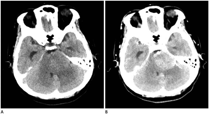
Fig. 1 Representative MRI of primary pontine glioblastoma multiforme in a 47-year-old female patient. A. Axial T2-weighted MR image demonstrates a heterogenously hyperintense tumor with partially defined margin. B. Axial pre-contrast T1-weighted MR image reveals a homogeneously hypointense tumor. C. Axial post-contrast fluid attenuated inversion recovery image shows a heterogeneously enhancing tumor with partial obstruction of the fourth ventricle and peritumoral vasogenic edema.
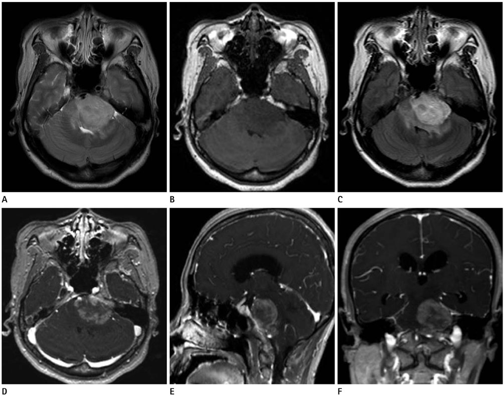
Fig. 2 Representative MRI of primary pontine glioblastoma multiforme in a 47-year-old female patient. A. Axial T2-weighted MR image demonstrates a heterogenously hyperintense tumor with partially defined margin. B. Axial pre-contrast T1-weighted MR image reveals a homogeneously hypointense tumor. C. Axial post-contrast fluid attenuated inversion recovery image shows a heterogeneously enhancing tumor with partial obstruction of the fourth ventricle and peritumoral vasogenic edema. D-F. Axial (D), sagittal (E), and coronal (F) enhanced T1-weighted MR images show a heterogeneously enhancing tumor with intratumoral necrosis involving pons and left Meckel's cave.
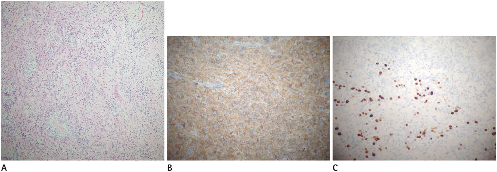
Fig. 3 Pathologic findings of primary pontine glioblastoma multiforme in a 47-year-old female patient. A. Photomicrograph (original magnification, × 100; hematoxylin and eosin stain) shows dense cellularity, nuclear pleomorphism and a variable nuclear:cytoplasmic ratio. B. GFAP staining (immunohistochemistry, × 200) shows diffusely positive immunoreactivity, presenting brownish staining. This result indicates astrocytic nature of tumor. C. Approximately 40% of cells have Ki-67 reactivity, which demonstrates increased cell proliferation (original magnification, × 200; Ki-67 stain). GFAP = glial fibrillary acidic protein
Reference
-
1. Guillamo JS, Monjour A, Taillandier L, Devaux B, Varlet P, Haie-Meder C, et al. Brainstem gliomas in adults: prognostic factors and classification. Brain. 2001; 124(Pt 12):2528–2539.2. Rachinger W, Grau S, Holtmannspötter M, Herms J, Tonn JC, Kreth FW. Serial stereotactic biopsy of brainstem lesions in adults improves diagnostic accuracy compared with MRI only. J Neurol Neurosurg Psychiatry. 2009; 80:1134–1139.3. Badhe PB, Chauhan PP, Mehta NK. Brainstem gliomas--a clinicopathological study of 45 cases with p53 immunohistochemistry. Indian J Cancer. 2004; 41:170–174.4. Theeler BJ, Ellezam B, Melguizo-Gavilanes I, de Groot JF, Mahajan A, Aldape KD, et al. Adult brainstem gliomas: correlation of clinical and molecular features. J Neurol Sci. 2015; 353:92–97.5. Reyes-Botero G, Mokhtari K, Martin-Duverneuil N, Delattre JY, Laigle-Donadey F. Adult brainstem gliomas. Oncologist. 2012; 17:388–397.6. Fischbein NJ, Prados MD, Wara W, Russo C, Edwards MS, Barkovich AJ. Radiologic classification of brain stem tumors: correlation of magnetic resonance imaging appearance with clinical outcome. Pediatr Neurosurg. 1996; 24:9–23.7. Lakhan SE, Harle L. Difficult diagnosis of brainstem glioblastoma multiforme in a woman: a case report and review of the literature. J Med Case Rep. 2009; 3:87.8. Kuroiwa T, Numaguchi Y, Rothman MI, Zoarski GH, Morikawa M, Zagardo MT, et al. Posterior fossa glioblastoma multiforme: MR findings. AJNR Am J Neuroradiol. 1995; 16:583–589.9. Zhang Y, Pan Y, Li X. Glioblastoma multiforme in the brainstem in a young adult. Clin Neurol Neurosurg. 2014; 124:175–178.10. Kwon JW, Kim IO, Cheon JE, Kim WS, Moon SG, Kim TJ, et al. Paediatric brain-stem gliomas: MRI, FDG-PET and histological grading correlation. Pediatr Radiol. 2006; 36:959–964.
- Full Text Links
-
- Actions
-
Cited
- CITED
-
- Close
- Share
-
- Similar articles
-
- Glioblastoma Multiforme with Subcutaneous Metastases, Case Report and Literature Review
- A Case of Multicentric Glioblastoma Multiforme
- A Case of Meningioma Compatible with Metastatic Glioblastoma Multiforme
- A Case of Glioblastoma Multiforme of the Cerebellum
- Scalp Metastasis of Glioblastoma Multiforme after Craniotomy and stereotatic Interstitial Brachytherapy
