Korean Circ J.
2016 May;46(3):408-411. 10.4070/kcj.2016.46.3.408.
Abdominal Wall Hematoma as a Rare Complication following Percutaneous Coronary Intervention
- Affiliations
-
- 1Division of Cardiology, Department of Internal Medicine, Hanyang University Guri Hospital, Hanyang University College of Medicine, Guri, Korea. cardio.hyapex@gmail.com
- 2Department of Radiology, Hanyang University Guri Hospital, Hanyang University College of Medicine, Guri, Korea.
- KMID: 2344454
- DOI: http://doi.org/10.4070/kcj.2016.46.3.408
Abstract
- Abdominal wall hematoma is a rare but potentially serious vascular complication that may develop after coronary angiographic procedures. In particular, an oblique muscle hematoma caused by an injury of the circumflex iliac artery is very rare, yet can be managed by conservative treatment including hydration and transfusion. However, when active bleeding continues, angiographic embolization or surgery might be needed. In this study, we report an uncommon case of injury to the circumflex iliac artery by an inappropriate introduction of the hydrophilic guidewire during the performance of a percutaneous coronary intervention.
MeSH Terms
Figure
-
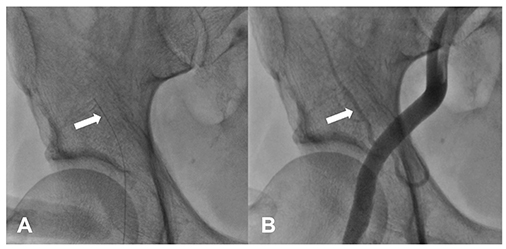
Fig. 1 Right femoral arteriography. (A) Hydrophilic guidewire is unintentionally introduced into the circumflex iliac artery (arrow). (B) Femoral and iliac arteriography after withdrawing the guidewire. The circumflex iliac artery is intact and there is no evidence of perforation (arrow).

Fig. 2 Abdomen computed tomography angiography. Transverse (A) and coronal (B) section show right-sided abdominal hematoma and circumflex iliac artery (arrows). There is no evidence of extravasation of contrast media.
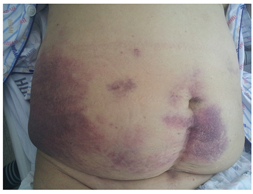
Fig. 3 Photograph of the patient with abdominal wall hematoma at 3 days after the procedure. Ecchymotic patches appears on the patient's lower abdomen, especially the right side.
Reference
-
1. Fukunaga N, Ikeyama S, Satomi J, Satoh K. Lateral abdominal wall hematoma as a rare complication after carotid artery stenting: a case report. World J Emerg Surg. 2009; 4:39.2. Merriweather N, Sulzbach-Hoke LM. Managing risk of complications at femoral vascular access sites in percutaneous coronary intervention. Crit Care Nurse. 2012; 32:16–29. quiz first page after 29.3. Hamel WJ. Femoral artery closure after cardiac catheterization. Crit Care Nurse. 2009; 29:39–46. quiz 47.4. Shimizu T, Hanasawa K, Yoshioka T, et al. Spontaneous hematoma of the lateral abdominal wall caused by a rupture of a deep circumflex iliac artery: report of two cases. Surg Today. 2003; 33:475–478.5. Ranieri P, Bianchetti A, Robecchi D, Trabucchi M. Spontaneous hematoma of the lateral abdominal wall: a case report. J Am Geriatr Soc. 2009; 57:2375–2376.6. Jabr FI, Skeik N. Spontaneous lateral abdominal wall hematoma complicating chronic obstructive pulmonary disease exacerbation. J Med Liban. 2011; 59:160–161.7. Luhmann A, Williams EV. Rectus sheath hematoma: a series of unfortunate events. World J Surg. 2006; 30:2050–2055.8. Dutta S, Sanjay P, Jones ML. Diagnosis and treatment of giant lateral abdominal wall haematoma after blunt trauma: a case report. Cases J. 2009; 2:9358.9. Shimodaira M, Kitano T, Kibata M, Shirahata K. An oblique muscle hematoma as a rare cause of severe abdominal pain: a case report. BMC Res Notes. 2013; 6:18.10. Fitts J, Ver Lee P, Hofmaster P, Malenka D; Northern New England Cardiovascular Study Group. Fluoroscopy-guided femoral artery puncture reduces the risk of PCI-related vascular complications. J Interv Cardiol. 2008; 21:273–278.11. Bangalore S, Bhatt DL. Femoral arterial access and closure. Circulation. 2011; 124:e147–e156.
- Full Text Links
-
- Actions
-
Cited
- CITED
-
- Close
- Share
-
- Similar articles
-
- Right Ventricular Wall Hematoma Secondary to Percutaneous Coronary Intervention
- Renal Subcapsular Hematoma after Percutaneous Transfemoral Angiography
- A Case of Right Coronary Artery Hematoma Detected by Echocardiography after Percutaneous Coronary Intervention
- A Case of Severe Cough-induced Abdominal Wall Hematoma
- Consecutive Multivessel Myocardial Infarction during Primary Percutaneous Coronary Intervention
