Radiologic and Pathologic Findings of a Follicular Variant of Papillary Thyroid Cancer with Extensive Stromal Fat: A Case Report
- Affiliations
-
- 1Department of Radiology, Konkuk University Medical Center, Konkuk University School of Medicine, Seoul 05030, Korea.
- 2Department of Pathology, Konkuk University Medical Center, Konkuk University School of Medicine, Seoul 05030, Korea.
- 3Department of Surgery, Konkuk University Medical Center, Konkuk University School of Medicine, Seoul 05030, Korea. ks2002p@hanmail.net
- KMID: 2344291
- DOI: http://doi.org/10.3348/kjr.2015.16.6.1349
Abstract
- Thyroid cancer may have small adipose structures detected by microscopy. However, there are no reports of thyroid cancer with gross fat evaluated by radiological methods. We reported a case of a 58-year-old woman with a fat containing thyroid mass. The mass was hyperechoic and ovoid in shape with a smooth margin on ultrasonography. On computed tomography, the mass had markedly low attenuation suggestive of fat, and fine reticular and thick septa-like structures. The patient underwent a right lobectomy. The mass was finally diagnosed as a follicular variant of papillary thyroid cancer with massive stromal fat.
MeSH Terms
Figure
-
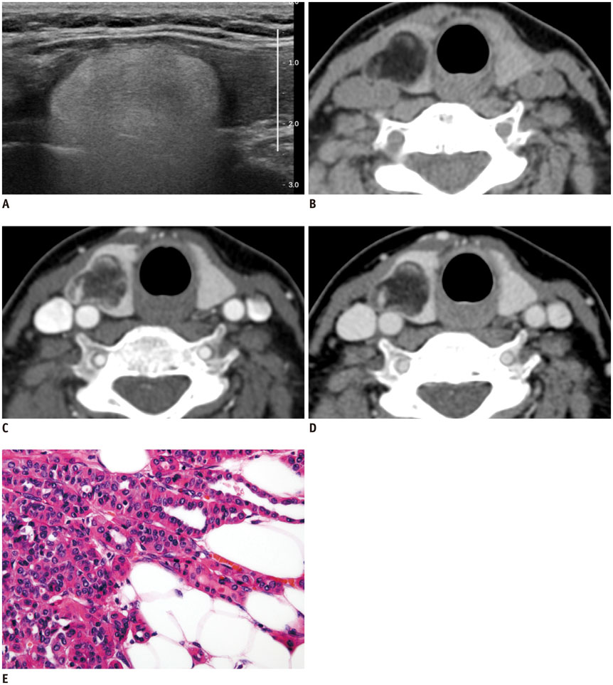
Fig. 1 58-year-old woman diagnosed with follicular variant of papillary thyroid cancer with mature fat. A. Longitudinal image of right thyroid using gray-scale ultrasonography shows hyperechoic, ovoid mass with smooth margin. Peripheral portion of mass is more echogenic than central area. Curtain-like hyperechoic shadowing was observed posterior to mass. Perithyroidal fat, muscle, and vertebral bodies were therefore not clearly visualized. B-D. Pre-contrast computed tomography (CT) shows fatty (mean CT number, -80 Hounsfield units, HU), well-defined mass in parenchyma of right mid-to-upper portion of thyroid (B). Mass has several fine reticular and thick septa-like soft tissue lesions, which show contrast enhancement. Post-contrast CT shows higher enhancement in early phase (C, at 40 seconds; mean CT number, 16 HU) of tumor than in delayed phase (D, 90 seconds; mean CT number, -13 HU). Tumor is lobular in appearance in axial plane (B-D), ovoid in longitudinal direction, and completely located in thyroid parenchyma without evidence of extrathyroidal extension. E. High-power magnification (1:400, hematoxylin and eosin staining) reveals tumor cells with enlarged nuclei, chromatin clearing, and nuclear grooves. Tumor cells are surrounded by mature adipose tissue.
Reference
-
1. Schröder S, Böcker W. Lipomatous lesions of the thyroid gland: a review. Appl Pathol. 1985; 3:140–149.2. Akslen LA, Maehle BO. Papillary thyroid carcinoma with lipomatous stroma. Am J Surg Pathol. 1997; 21:1256–1257.3. Azar AR, Weynand B, Daumerie C, Coche E. Metastatic liposarcoma of the thyroid gland. Br J Radiol. 2003; 76:750–752.4. Bisi H, Longatto Filho A, de Camargo RY, Fernandes VS. Thyroid papillary carcinoma lipomatous type: report of two cases. Pathologica. 1993; 85:761–764.5. DeRienzo D, Truong L. Thyroid neoplasms containing mature fat: a report of two cases and review of the literature. Mod Pathol. 1989; 2:506–510.6. Vestfrid MA. Papillary carcinoma of the thyroid gland with lipomatous stroma: report of a peculiar histological type of thyroid tumour. Histopathology. 1986; 10:97–100.7. Zhang YZ, Li WH, Zhu MJ, Li YH, Gao Y. An unusual mature thyroid teratoma on CT and 99Tcm scintigraphy imaging in a child. Pediatr Radiol. 2010; 40:1831–1833.8. Demirpolat G, Guney B, Savas R, Tuncay G, Alper H, Sener RN. Radiologic and cytologic findings in a case of thyrolipoma. AJNR Am J Neuroradiol. 2002; 23:1640–1641.9. Kim KH, Seo HS, Lee YH, Lee KY, Kim YS, Son GS, et al. Study of intrathyroid fat-containing lesions using CT imaging with literature review. Neuroradiology. 2013; 55:1405–1411.10. Ge Y, Luna MA, Cowan DF, Truong LD, Ayala AG. Thyrolipoma and thyrolipomatosis: 5 case reports and historical review of the literature. Ann Diagn Pathol. 2009; 13:384–389.11. Daboin KP, Ochoa-Perez V, Luna MA. Adenolipomas of the head and neck: analysis of 6 cases. Ann Diagn Pathol. 2006; 10:72–76.12. Borges A, Catarino A. Case 53: adenolipoma of the thyroid gland. Radiology. 2002; 225:746–750.
- Full Text Links
-
- Actions
-
Cited
- CITED
-
- Close
- Share
-
- Similar articles
-
- Differential Diagnosis of a Follicular Carcinoma and Papillary Carcinoma of the Thyroid Gland Based on Sonographic Findings
- Demonstration of TCM-9 Monoclonal Antibody in Follicular Neoplasm of Thyroid
- Pathologic basis of the sonographic differences between thyroid cancer and noninvasive follicular thyroid neoplasm with papillary-like nuclear features
- Macrofollicular Variant of Papillary Thyroid Carcinoma with Extensive Hemorrhage
- Diagnostic Dilemma of a Follicular Lesions/Neoplasm in Thyroid Fine Needle Aspiration Cytology
