Korean J Ophthalmol.
2014 Oct;28(5):359-363. 10.3341/kjo.2014.28.5.359.
Laser-assisted In Situ Keratomileusis for Correction of Astigmatism and Increasing Contact Lens Tolerance after Penetrating Keratoplasty
- Affiliations
-
- 1Department of Ophthalmology, Uijeongbu St. Mary's Hospital, The Catholic University of Korea College of Medicine, Uijeongbu, Korea.
- 2Hospital of 18th Fighter Wing, Republic of Korea Air Force, Korea.
- 3Department of Ophthalmology, Seoul St. Mary's Hospital, The Catholic University of Korea College of Medicine, Seoul, Korea. mskim@catholic.ac.kr
- KMID: 2344258
- DOI: http://doi.org/10.3341/kjo.2014.28.5.359
Abstract
- PURPOSE
To determine effectiveness of laser-assisted in situ keratomileusis (LASIK) in the treatment of astigmatism following penetrating keratoplasty (PK).
METHODS
We performed a retrospective review of medical records of patients who underwent LASIK following PK and had over 1 year of follow-up data.
RESULTS
Twenty-six patients (26 pairs of eyes) underwent LASIK following PK. Mean age of the patients at the time of LASIK was 40.7 years (range, 26 to 72 years). Following LASIK, the mean cylinder was reduced by 2.4 diopters and mean reduction of cylinder after LASIK was 65.4% from the preoperative values at the last follow-up visit. Uncorrected visual acuity became 20 / 50 or better in 69.2% of the eyes after LASIK. Best-corrected visual acuity became 20 / 50 or better in 73.1% of the eyes after LASIK. All of them were intolerable to contact lenses before LASIK. After LASIK, 6 pairs (23.1%) did not need to use contact lenses and 18 pairs (69.2%) were tolerable to using contact lenses or spectacles. There were no significant endothelial cell density changes 12 months after LASIK (p = 0.239).
CONCLUSIONS
LASIK is effective in the treatment of astigmatism following PK and increases contact lens and spectacle tolerance.
MeSH Terms
-
Adult
Aged
Astigmatism/etiology/physiopathology/*surgery
*Contact Lenses/utilization
Corneal Topography
Female
Humans
Keratomileusis, Laser In Situ/*methods
Keratoplasty, Penetrating/*adverse effects
Lasers, Excimer/*therapeutic use
Male
Middle Aged
Refraction, Ocular/physiology
Retrospective Studies
Vision, Binocular/physiology
Visual Acuity/physiology
Figure
-
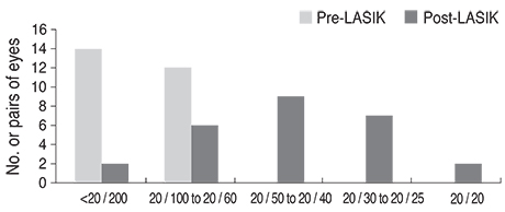
Fig. 1 Uncorrected visual acuity before and after laser-assisted in situ keratomileusis (LASIK).

Fig. 2 Change in uncorrected visual acuity (UCVA) after laserassisted in situ keratomileusis.
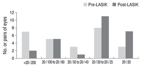
Fig. 3 Best-corrected visual acuity before and after laser-assisted in situ keratomileusis (LASIK).

Fig. 4 Change in best corrected visual acuity (BCVA) after laserassisted in situ keratomileusis.

Fig. 5 Comparison of mean cylinder in diopters before and after laser-assisted in situ keratomileusis (LASIK).

Fig. 6 Contact lens or spectacle tolerance before and after laserassisted in situ keratomileusis (LASIK).
Reference
-
1. Lazzaro DR, Haight DH, Belmont SC, et al. Excimer laser keratectomy for astigmatism occurring after penetrating keratoplasty. Ophthalmology. 1996; 103:458–464.2. Bansal AK. Photoastigmatic refractive keratectomy for correction of astigmatism after keratoplasty. J Refract Surg. 1999; 15:2 Suppl. S243–S245.3. Campos M, Hertzog L, Garbus J, et al. Photorefractive keratectomy for severe postkeratoplasty astigmatism. Am J Ophthalmol. 1992; 114:429–436.4. Bilgihan K, Ozdek SC, Akata F, Hasanreisoglu B. Photorefractive keratectomy for post-penetrating keratoplasty myopia and astigmatism. J Cataract Refract Surg. 2000; 26:1590–1595.5. Chan WK, Hunt KE, Glasgow BJ, Mondino BJ. Corneal scarring after photorefractive keratectomy in a penetrating keratoplasty. Am J Ophthalmol. 1996; 121:570–571.6. Genvert GI, Cohen EJ, Arentsen JJ, Laibson PR. Fitting gas-permeable contact lenses after penetrating keratoplasty. Am J Ophthalmol. 1985; 99:511–514.7. Koffler BH, Clements LD, Litteral GL, Smith VM. A new contact lens design for post-keratoplasty patients. CLAO J. 1994; 20:170–175.8. Matsuda M, MacRae SM, Inaba M, Manabe R. The effect of hard contact lens wear on the keratoconic corneal endothelium after penetrating keratoplasty. Am J Ophthalmol. 1989; 107:246–251.9. Kohnen T, Buhren J. Corneal first-surface aberration analysis of the biomechanical effects of astigmatic keratotomy and a microkeratome cut after penetrating keratoplasty. J Cataract Refract Surg. 2005; 31:185–189.10. Alio JL, Javaloy J, Osman AA, et al. Laser in situ keratomileusis to correct post-keratoplasty astigmatism: 1-step versus 2-step procedure. J Cataract Refract Surg. 2004; 30:2303–2310.11. Barraquer CC, Rodriguez-Barraquer T. Five-year results of laser in-situ keratomileusis (LASIK) after penetrating keratoplasty. Cornea. 2004; 23:243–248.12. Buzard K, Febbraro JL, Fundingsland BR. Laser in situ keratomileusis for the correction of residual ametropia after penetrating keratoplasty. J Cataract Refract Surg. 2004; 30:1006–1013.13. Hardten DR, Chittcharus A, Lindstrom RL. Long term analysis of LASIK for the correction of refractive errors after penetrating keratoplasty. Cornea. 2004; 23:479–489.14. Ranchod TM, McLeod SD. Wound dehiscence in a patient with keratoconus after penetrating keratoplasty and LASIK. Arch Ophthalmol. 2004; 122:920–921.15. Dawson DG, Hardten DR, Albert DM. Pocket of fluid in the lamellar interface after penetrating keratoplasty and laser in situ keratomileusis. Arch Ophthalmol. 2003; 121:894–896.
- Full Text Links
-
- Actions
-
Cited
- CITED
-
- Close
- Share
-
- Similar articles
-
- Three Cases of LASIK for Myopia and Astigmatism after Penetrating Keratoplasty
- The Correction of Corneal Astigmatism Using Piggyback Contact Lens
- Surgical Correction of Hyperopia
- The Effect of PRK and LASIK for the Correction of Postkeratoplasty Astigmatism
- Ocular deviation after unilateral laser in situ keratomileusis
