A Korean Woman with Reactive Lymphoid Hyperplasia of the Uvea
- Affiliations
-
- 1Institute of Vision Research, Department of Ophthalmology, International St. Mary's Hospital, Yonsei University College of Medicine, Incheon, Korea.
- 2Institute of Vision Research, Department of Ophthalmology, Yonsei University College of Medicine, Seoul, Korea. sunglee@yuhs.ac
Abstract
- No abstract available.
MeSH Terms
-
Asian Continental Ancestry Group/ethnology
Choroid Diseases/*diagnosis/drug therapy/ethnology
Female
Fluorescein Angiography
Glucocorticoids/therapeutic use
Humans
Middle Aged
Multimodal Imaging
Ophthalmoscopy
Prednisolone/therapeutic use
Pseudolymphoma/*diagnosis/drug therapy/ethnology
Republic of Korea/epidemiology
Tomography, Optical Coherence
Ultrasonography
Glucocorticoids
Prednisolone
Figure
-
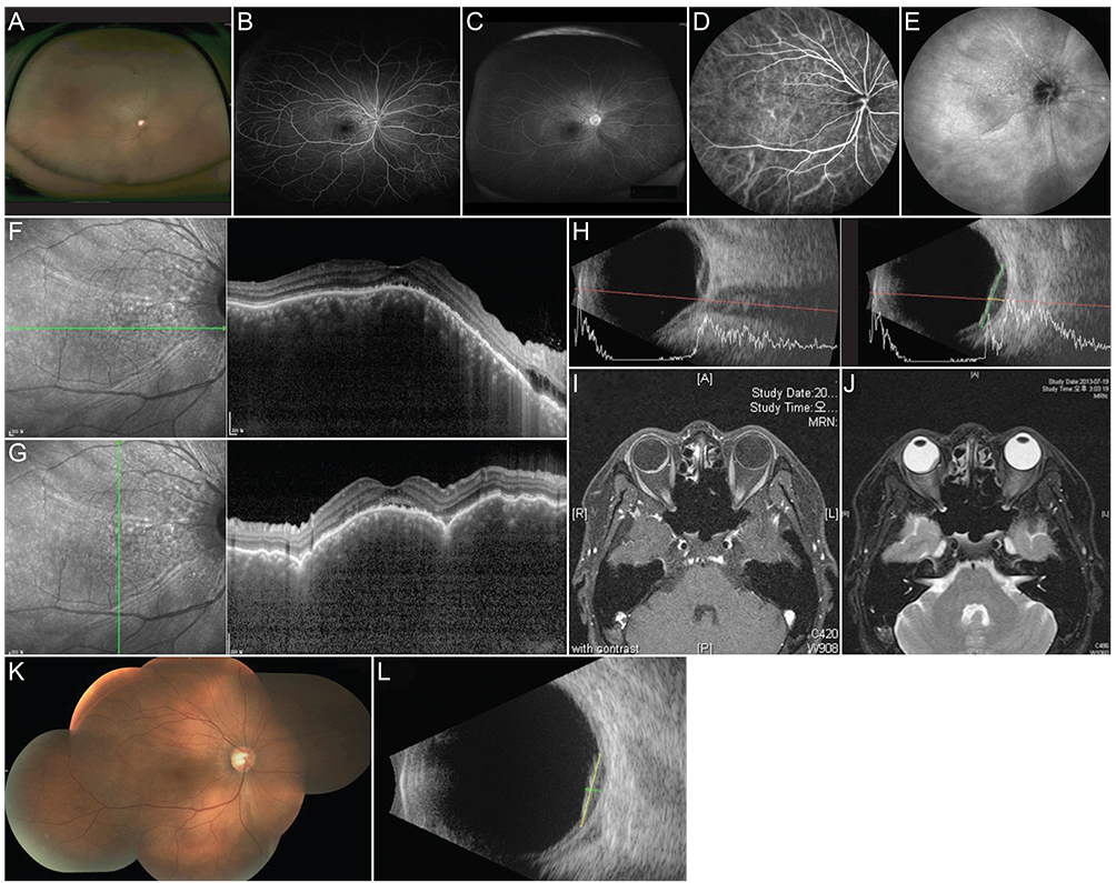
Fig. 1 Fundus photography of the 50-year-old patient with reactive lymphoid hyperplasia of the uvea in the right eye (A). Fluorescence angiography showed stippled hyperfluorescence (B) with late phase staining (C). On indocyanine green angiography, retinochoroidal folds radiating from the optic nerve head were seen as hypofluorescence lines throughout examination (D,E). Spectral domain optical coherence tomography showed subretinal fluid at fovea, with undulating retina due to choroidal mass both horizontally (F) and vertically (G). Ultrasound sonography showed homogenous intraocular elevation which was acoustically empty (H), and lesion size was base 11.93 mm with height 2.71 mm. On magnetic resonance imaging of brain and orbit, the subretinal mass showed hyperintensity in T1 (I), and hypointesnity on T2 images (J). After systemic treatment with steroid for 2 weeks, choroidal elevation was improved (K). On ultrasonography, size of choroidal mass was measured as base 8.80 mm with height 1.87 mm (L).
Reference
-
1. Gass JD. Retinal detachment and narrow-angle glaucoma secondary to inflammatory pseudotumor of the uveal tract. Am J Ophthalmol. 1967; 64:Suppl. 612–621.2. Ryan SJ, Zimmerman LE, King FM. Reactive lymphoid hyperplasia. An unusual form of intraocular pseudotumor. Trans Am Acad Ophthalmol Otolaryngol. 1972; 76:652–671.3. Grossniklaus HE, Martin DF, Avery R, et al. Uveal lymphoid infiltration: report of four cases and clinicopathologic review. Ophthalmology. 1998; 105:1265–1273.4. Desroches G, Abrams GW, Gass JD. Reactive lymphoid hyperplasia of the uvea: a case with ultrasonographic and computed tomographic studies. Arch Ophthalmol. 1983; 101:725–728.5. Chang TS, Byrne SF, Gass JD, et al. Echographic findings in benign reactive lymphoid hyperplasia of the choroid. Arch Ophthalmol. 1996; 114:669–675.
- Full Text Links
-
- Actions
-
Cited
- CITED
-
- Close
- Share
-
- Similar articles
-
- Orbital Lymphocytic Tumor Gradually Progressed to More Malignant Form during 4 Years
- Diagnostic Pediatric Colonoscopy for Lymphoid Hyperplasia of Terminal Ileum
- Reactive Lymphoid Hyperplasia Treated with Radiofrequency Ablation
- A Case of Ocular Adnexal Benign Reactive Lymphoid Hyperplasia Recurred as Systemic Malignant Lymphoma
- Two Cases of Small Intestinal Nodular Lymphoid Hyperplasia
