Korean J Ophthalmol.
2014 Aug;28(4):330-336.
Patterns of Subsequent Progression of Localized Retinal Nerve Fiber Layer Defects on Red-free Fundus Photographs in Normal-tension Glaucoma
- Affiliations
-
- 1Department of Ophthalmology, Seoul National University Hospital, Seoul, Korea. dmkim@snu.ac.kr
Abstract
- PURPOSE
To investigate patterns of subsequent progression of localized retinal nerve fiber layer (RNFL) defects and to quantify the extent of progression in normal-tension glaucoma (NTG) patients.
METHODS
Thirty-three eyes of 33 consecutive NTG patients who had shown continuous progression of localized RNFL defect on serial red-free fundus photographs were selected for the study. Patterns of subsequent progression of localized RNFL defects were categorized, and extents of progression were quantified. Serial evaluations of disc stereophotographs and visual fields were also performed to detect progression.
RESULTS
The most common pattern was continuous widening of the defect towards the macula (n = 11, 33.3%) followed by sharpening of the defect border after widening of the defect towards the macula (n = 5, 15.2%), continuous widening of the defect away from the macula (n = 2, 6.1%), and deepening of the defect after appearance of a new defect (n = 2, 6.1%). Four eyes (12.1%) simultaneously showed two patterns of subsequent progression. In 13 eyes that showed continuous widening of the defect, subsequent angular widening towards the macula and away from the macula were 9.2 ± 6.0degrees (range, 1.1degrees to 24.4degrees; n = 11) and 5.2 ± 4.9degrees (range, 0.3degrees to 11.3degrees; n = 2), respectively. Thirty-two eyes showed no progression of optic disc cupping. Out of the 21 eyes in which Humphrey central 30-2 threshold visual field tests were performed after progression of RNFL defects, 15 eyes showed no deterioration in the visual field.
CONCLUSIONS
There were nine patterns of subsequent progression of localized RNFL defects. Among them, continuous RNFL loss proceeding temporally was the most common one. Initial progression of the defect proceeded temporally, especially in the defect located at the inferior fundus, might be at a risk of further RNFL loss temporally.
Keyword
MeSH Terms
Figure
-
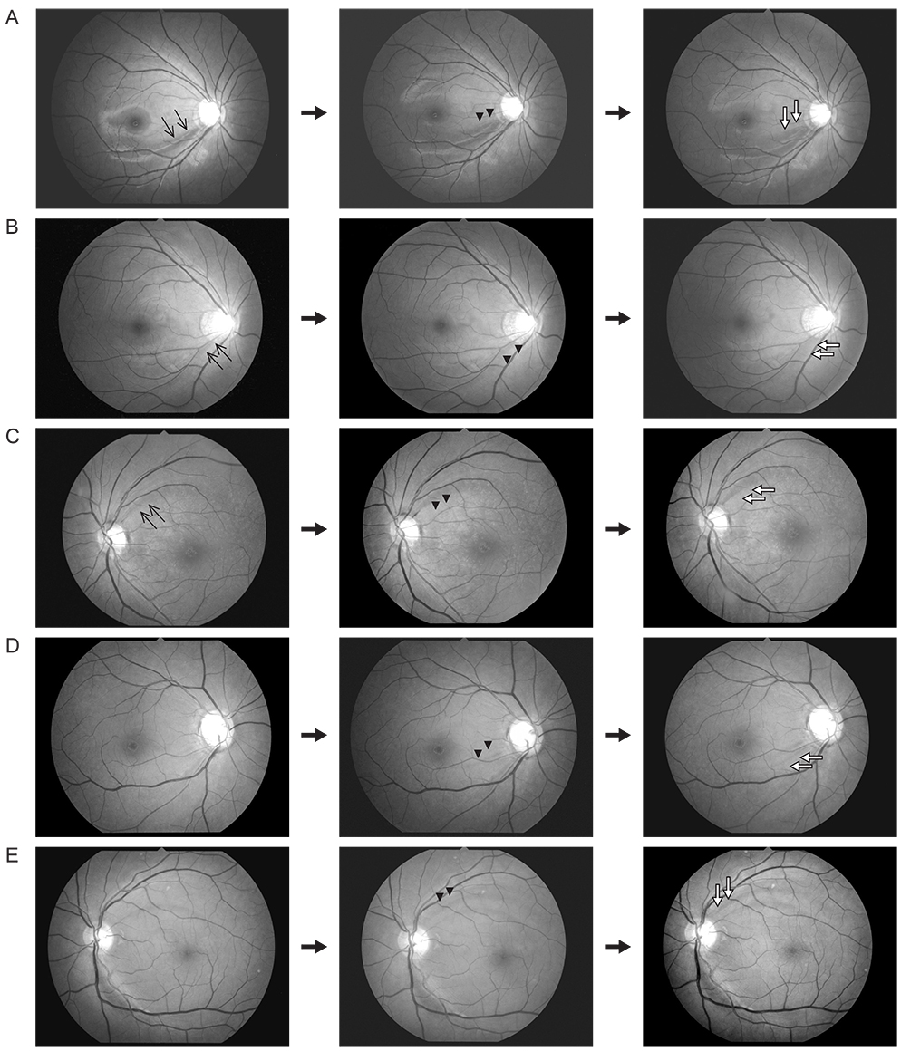
Fig. 1 Red-free fundus photographs show patterns of continuous progression of localized retinal nerve fiber layer defects such as (A) continuous widening (arrow heads and empty arrow) of the defect towards the macula, (B) continuous widening (arrow heads and empty arrow) of the defect away from the macula, (C) continuous sharpening of the border (arrow heads and empty arrow) of the defect, (D) continuous appearance of a new defect (arrow heads and empty arrow), and (E) appearance of a new defect (arrow heads) followed by deepening of the defect (empty arrow). Arrows in the left first photographs indicate the location of the defect.
Reference
-
1. Tuulonen A, Airaksinen PJ. Initial glaucomatous optic disk and retinal nerve fiber layer abnormalities and their progression. Am J Ophthalmol. 1991; 111:485–490.2. Quigley HA, Reacher M, Katz J, et al. Quantitative grading of nerve fiber layer photographs. Ophthalmology. 1993; 100:1800–1807.3. Sommer A, Miller NR, Pollack I, et al. The nerve fiber layer in the diagnosis of glaucoma. Arch Ophthalmol. 1977; 95:2149–2156.4. Quigley HA, Katz J, Derick RJ, et al. An evaluation of optic disc and nerve fiber layer examinations in monitoring progression of early glaucoma damage. Ophthalmology. 1992; 99:19–28.5. Kim KE, Ahn SJ, Kim DM. Comparison of two different spectral domain optical coherence tomography devices in the detection of localized retinal nerve fiber layer defects. Jpn J Ophthalmol. 2013; 57:347–358.6. Jeoung JW, Kim TW, Weinreb RN, et al. Diagnostic ability of spectral-domain versus time-domain optical coherence tomography in preperimetric glaucoma. J Glaucoma. 2013; 01. 31. http://dx.doi.org/10.1097/IJG.0b013e3182741cc4.7. Nukada M, Hangai M, Mori S, et al. Detection of localized retinal nerve fiber layer defects in glaucoma using enhanced spectral-domain optical coherence tomography. Ophthalmology. 2011; 118:1038–1048.8. Suh MH, Kim DM, Kim YK, et al. Patterns of progression of localized retinal nerve fibre layer defect on red-free fundus photographs in normal-tension glaucoma. Eye (Lond). 2010; 24:857–863.9. Hoyt WF, Frisen L, Newman NM. Fundoscopy of nerve fiber layer defects in glaucoma. Invest Ophthalmol. 1973; 12:814–829.10. Boden C, Blumenthal EZ, Pascual J, et al. Patterns of glaucomatous visual field progression identified by three progression criteria. Am J Ophthalmol. 2004; 138:1029–1036.11. Jonas JB, Schiro D. Localised wedge shaped defects of the retinal nerve fibre layer in glaucoma. Br J Ophthalmol. 1994; 78:285–290.12. Ishida K, Yamamoto T, Sugiyama K, Kitazawa Y. Disk hemorrhage is a significantly negative prognostic factor in normal-tension glaucoma. Am J Ophthalmol. 2000; 129:707–714.13. Airaksinen PJ, Tuulonen A, Werner EB. Clinical evaluation of the optic disc and retinal nerve fiber layer. In : Ritch R, Shields MB, Krupin T, editors. The glaucomas. 2nd ed. St Louis: Mosby;1996. p. 617–657.14. Kim NR, Lee ES, Seong GJ, et al. Spectral-domain optical coherence tomography for detection of localized retinal nerve fiber layer defects in patients with open-angle glaucoma. Arch Ophthalmol. 2010; 128:1121–1128.
- Full Text Links
-
- Actions
-
Cited
- CITED
-
- Close
- Share
-
- Similar articles
-
- The Detection of Retinal Never Fiber Layer Defect by Modification of Non-mydriatic Digital Fundus Photograph
- Scanning Laser Polarimetry and Optical Coherence Tomography for Detection of Retinal Nerve Fiber Layer Defects
- Early Detection of Glaucomatous Optic Nerve Abnormality by Nonmydriatic Digital Fundus Camera in a Routine Health Check-up
- Progression of Glaucoma in Highly Myopic Eyes with Paravascular Inner Retinal Defects
- Usefulness of Automated Measurements of Localized Retinal Nerve Fiber Layer Defects Area Using Significance Map
