J Korean Ophthalmol Soc.
2014 Aug;55(8):1238-1241.
Posterior Polymorphous Corneal Dystrophy (PPCD) Combined with Traumatic Descemet's Membrane Fold
- Affiliations
-
- 1Department of Ophthalmology and Visual Science, Seoul St. Mary's Hospital, The Catholic University of Korea College of Medicine, Seoul, Korea. mskim@catholic.ac.kr
Abstract
- PURPOSE
To present a case of a 72-year-old woman with posterior polymorphous corneal dystrophy (PPCD) combined with traumatic Descemet's membrane fold due to forceps injury in Birth.
CASE SUMMARY
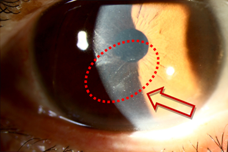
A 72-year-old woman without significant ocular history presented complaining of ocular discomfort in her right eye. She had a history of birth that required forceps delivery and poor vision in the right eye since childhood. The rest of perinatal ophthalmic history was unremarkable (no past ophthalmic or family history). The best corrected visual acuity was 20/20 in both eyes. Slit-lamp biomicroscopy showed distinctive, vertical, semitranslucent Descemet's membrane breaks situated on the superior temporal side of the posterior surface of the cornea in her right eye. Also, a horizontal, tram-track line appearance of posterior cornea surface was detected at middle and inferior side of cornea. The fellow eye was normal. The endothelial cell densities were 945 cells/mm2 and 2481 cells/mm2 in right and left eyes, respectively. After 11 years later, (routine follow up exams every 1 year), the endothelial cell densities were 901 cells/mm2 and 2481 cells/mm2 in right and left eyes, respectively, which means there were no significant changes of endothelial densities in both eyes.
CONCLUSIONS
We report a case of a patient with posterior polymorphous corneal dystrophy (PPCD) combined with traumatic Descemet's membrane fold due to forceps injury in Birth. The disease does not seem to progress or aggravated in long term follow up and no specific treatment was required.
Keyword
MeSH Terms
Figure
Reference
-
References
1. Kaufman HE, Barron BA, McDonald MB. The cornea. 1st ed.New York: Churchill Livingstone;1988. p. 432–8.2. Cibis GW, Krachmer JA, Phelps CD, Weingeist TA. The clinical spectrum of posterior polymorphous dystrophy. Arch Ophthalmol. 1977; 95:1529–37.
Article3. Grayson M. The nature of hereditary deep polymorphous dystrophy of the cornea: its association with iris and anterior chamber dygenesis. Trans Am Ophthalmol Soc. 1974; 72:516–59.4. Park IC, Chung SK, Myong YW, Rhee SW. A case of posterior polymorphous dystrophy. J Korean Ophthalmol Soc. 1991; 32:1020–3.5. Krachmer JH, Mannis MJ, Holland EJ. Cornea. 2nd ed.1. Philadelphia, PA: Elsevier Mosby;2005.6. Koeppe L. Klinische Beobachtungen mit der Nernst Spaltlampe und dem Hornhautmikroskop. Albrecht von Graefes Arch Ophthalmol. 1916; 91:375–9.7. Babu K, Murthy KR. In vivo confocal microscopy in different types of posterior polymorphous dystrophy. Indian J Ophthalmol. 2007; 55:376–8.8. Patel DV, Grupcheva CN, McGhee CN. In vivo confocal microscopy of posterior polymorphous dystrophy. Cornea. 2005; 24:550–4.
Article9. Hofmann RF, Paul TO, Pentelei-Molnar J. The management of corneal birth trauma. J Pediatr Ophthalmol Strabismus. 1981; 18:45–7.
Article10. Lisch W. Corneal lesion by vacuum extraction (author's transl). Klin Monbl Augenheilkd. 1976; 169:520–3.
- Full Text Links
- Actions
-
Cited
- CITED
-
- Close
- Share
- Similar articles
-
- A Case of Posterior Polymorphous Dystrophy
- Confocal Microscopic Findings in Posterior Polymorphous Corneal Dystrophy
- The Clinical Features and Progression of the Disease in Posterior Polymorphous Corneal Dystrophy (PPCD)
- Histological Characteristics of the Interface of Corneal Stroma and Descemet's Membrane
- Confocal Microscopic Findings of Corneal Tissue in Fuchs' Corneal Endothelial Dystrophy


