J Korean Ophthalmol Soc.
2014 Apr;55(4):628-632.
Bilateral Optic Neuropathy in Middle-Aged Woman Associated with Charcot Marie Tooth Disease Type 2A: A Case Report
- Affiliations
-
- 1Department of Ophthalmology, Seoul National University College of Medicine, Seoul, Korea. ophjun@gmail.com
- 2Seoul Artificial Eye Center, Seoul National University Hospital Clinical Research Institute, Seoul, Korea.
Abstract
- PURPOSE
Charcot-Marie-Tooth disease type 2A (CMT2A) is caused by mutations in the mitofusin 2 (MFN2) genes associated with variable central nervous system (CNS) involvement. The authors report a case of a middle-aged woman with genetically confirmed CMT type 2 (CMT2), combined with delayed-onset bilateral optic neuropathy.
CASE SUMMARY
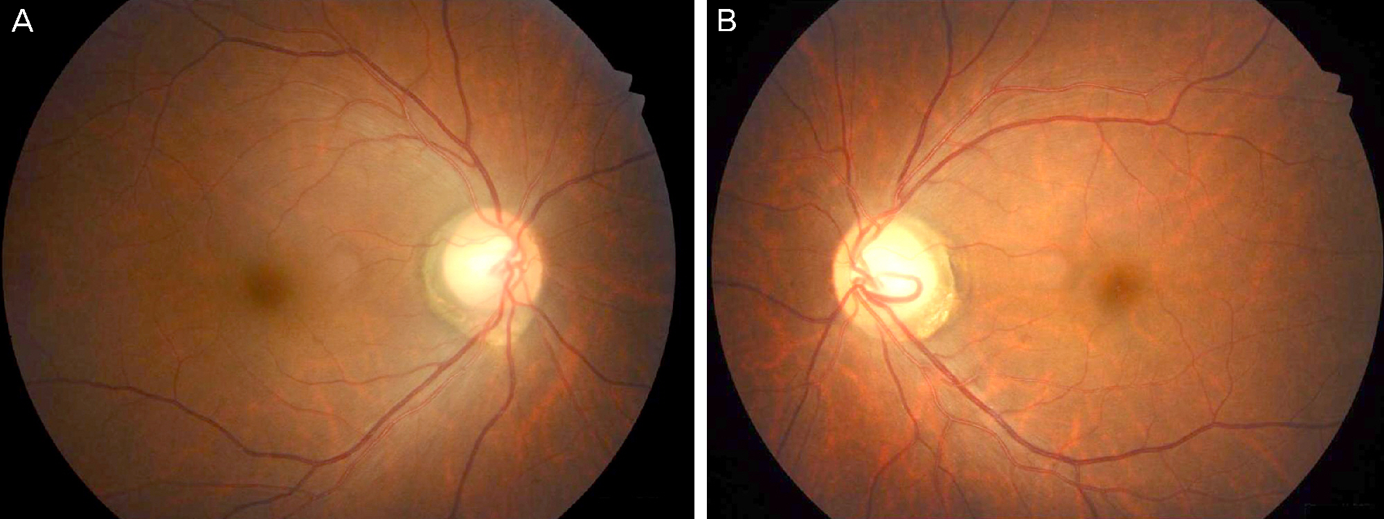
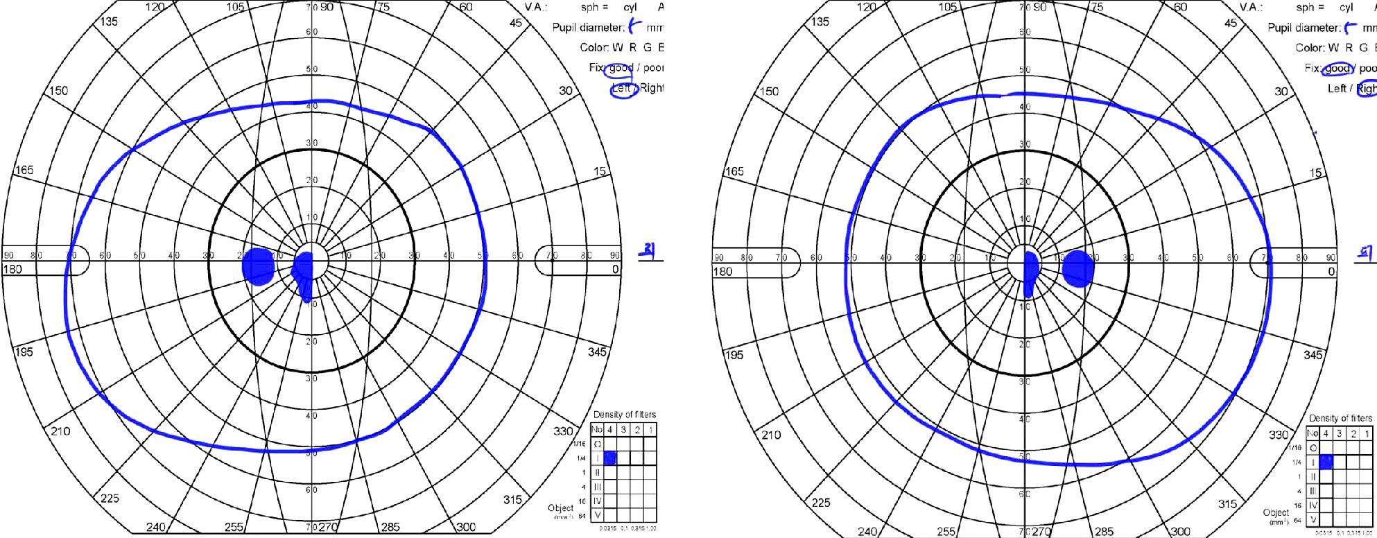
A 47-year-old woman presented with complaints of subacute decrease of visual acuity in both eyes. Her corrected visual acuity was 20/200 in the right eye and 20/320 in the left eye. Fundus photographs revealed bilateral disc pallor and diffuse retinal nerve fiber layer defects. No papillomacular bundle defect was observed. Goldmann perimetry showed central scotoma in both eyes. She had suffered from muscle wasting of the legs and foot deformities such as high arches and hammer toes since childhood and required a wheelchair for ambulation. A series of CMT gene mutation tests revealed an MFN2 gene mutation, c.617C>T (p.Thr206Ile), and the patient was diagnosed with CMT2A.
CONCLUSIONS
Charcot-Marie-Tooth disease is a common inherited neuromuscular disorder and CMT2A, an axonal CMT neuropathy, is associated with bilateral optic neuropathy. Therefore, suspecting CMT and testing for gene mutations as part of the work-up in patients with subacute bilateral optic neuropathy associated with peripheral neuropathy is critical.
MeSH Terms
Figure
Reference
-
References
1. Voo I, Allf BE, Udar N, et al. Hereditary motor and sensory neuropathy type VI with optic atrophy. Am J Ophthalmol. 2003; 136:670–7.
Article2. Shy ME. Charcot-Marie-Tooth disease: an update. Curr Opin Neurol. 2004; 17:579–85.
Article3. Skre H. Genetic and clinical aspects of Charcot-Marie-Tooth's disease. Clin Genet. 1974; 6:98–118.
Article4. Pareyson D, Marchesi C. Diagnosis, natural history, and management of Charcot-Marie-Tooth disease. Lancet Neurol. 2009; 8:654–67.
Article5. Barisic N, Claeys KG, Sirotkovic-Skerlev M, et al. Charcot-Marie-Tooth disease: a clinico-genetic confrontation. Ann Hum Genet. 2008; 72:416–41.
Article6. Zühner S, De Jonghe P, Jordanova A, et al. Axonal neuropathy with optic atrophy is caused by mutations in mitofusin 2. Ann Neurol. 2006; 59:276–81.7. Chung KW, Kim SB, Park KD, et al. Early onset severe and late-onset mild Charcot-Marie-Tooth disease with mitofusin 2 (MFN2) mutations. Brain. 2006; 129:2103–18.
Article8. Wakerley BR, Harman FE, Altmann DM, Malik O. Charcot-Marie-Tooth disease associated with recurrent optic neuritis. J Clin Neurosci. 2011; 18:1422–3.
Article9. Kim HJ, Sohn KM, Shy ME, et al. Mutations in PRPS1, which enc-odes the phosphoribosyl pyrophosphate synthetase enzyme critical for nucleotide biosynthesis, cause hereditary peripheral neuropathy with hearing loss and optic neuropathy (cmtx5). Am J Hum Genet. 2007; 81:552–8.
Article10. Sadun AA, Win PH, Ross-Cisneros FN, et al. Leber's hereditary optic neuropathy differentially affects smaller axons in the optic nerve. Trans Am Ophthalmol Soc. 2000; 98:223–32.
- Full Text Links
- Actions
-
Cited
- CITED
-
- Close
- Share
- Similar articles
-
- Hereditary Motor and Sensory Neuropathy Type VI with Bilateral Middle Cerebellar Peduncle Involvement
- A novel p.Leu699Pro mutation in MFN2 gene causes Charcot-Marie-Tooth disease type 2A
- A Case Report of Neuronal Type of Charcot-Marie-Tooth Disease
- A Family of Hereditary Neuropathy with Liability to Pressure Palsy Presenting Atypical Electrophysiological Features
- DNA diagnostic testing in hereditary motor and sensory neuropathies


