A Case of Primary Signet Ring Cell Carcinoma of the Lower Eyelid
- Affiliations
-
- 1Department of Ophthalmology, Wonkwang University School of Medicine, Institute of Wonkwang Medical Science, Iksan, Korea. sangduck@wonkwang.ac.kr
- 2Department of Pathology, Wonkwang University School of Medicine, Institute of Wonkwang Medical Science, Iksan, Korea.
Abstract
- PURPOSE
To report a rare case of a painless mass on the lower lid, histologically diagnosed as primary signet ring cell carcinoma of the eyelid.
CASE SUMMARY
A 74-year-old male presented with a painless mass on the right lower lid which had developed seven months prior to presentation. Incisional biopsy of the mass and attached lower lid skin was performed, revealing signet ring cell carcinoma. A systemic evaluation, including whole body positron emission tomography-computed tomography (PET-CT), chest CT, abdomen and pelvis CT, gastrointestinal endoscopy, and colonoscopy, revealed no other abnormal lesions. Therefore, the eyelid lesion was considered primary signet ring cell carcinoma of the skin and was treated with radiotherapy of 6600 cGy in 33 fractions over 7 weeks.
CONCLUSIONS
Herein, the authors report a rare case of primary signet ring cell carcinoma of the eyelid with no evidence of tumor recurrence or metastasis for 5 years after radiotherapy.
Keyword
MeSH Terms
Figure
-

Figure 1. Clinical photograph of the painless mass (black arrow) of the right lower eyelid.
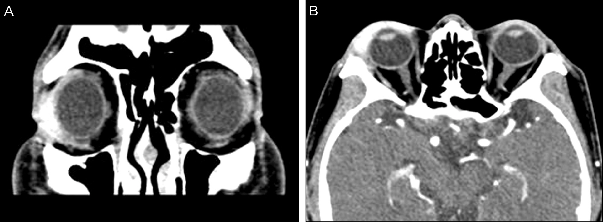
Figure 2. Coronal (A) and axial (B) images of orbital computed tomography scan showing a mass in the right inferolateral orbit and diffuse soft tissue thickening with well enhancement in the right lacrimal fossa and preseptal area.

Figure 3. (A, B) Histopathology reveals the tumor cells have ec-centric nuclei and some of the tumor cells show signet ring-like appearance (black arrow) in the fibrosclerotic background (A: H&E, ×400) and in the skeletal muscle fibers (B: H&E, ×200).(C) The tumor cells show positive staining for cytokeratin (×200).

Figure 4. Orbital magnetic resonance imaging (MRI) showing no evidence of recurrence 5 years after radiotherapy. Post contrast T1-weighted axial (A) and coronal (B) views.
Reference
-
References
1. Rosen Y, Kim B, Yermakov VA. Eccrine sweat gland tumor of clear cell origin involving the eyelids. Cancer. 1975; 36:1034–41.
Article2. Tanboon J, Uiprasertkul M, Luemsamran P. Signet-ring cell/histiocytoid carcinoma of the eyelid: a case report and review of the literature. Am J Dermatopathol. 2013; 35:e1–5.3. Wollensak G, Witschel H, Böhm N. Signet ring cell carcinoma of the eccrine sweat glands in the eyelid. Ophthalmology. 1996; 103:1788–93.
Article4. Requena L, Prieto VG, Requena C, et al. Primary signet-ring cell/histiocytoid carcinoma of the eyelid: a clinicopathologic study of 5 cases and review of the literature. Am J Surg Pathol. 2011; 35:378–91.5. Mortensen AL, Heegaard S, Clemmensen O, Prause JU. Signet ring cell carcinoma of the eyelid-the monocle tumor. APMIS. 2008; 116:326–32.6. Iwaya M, Uehara T, Yoshizawa A, et al. A case of primary signet-ring cell/histiocytoid carcinoma of the eyelid: immunohistochemical comprasion with the normal sweat gland and review of the literature. Am J Dermatopathol. 2012; 34:e139–45.7. Langel DJ, Yeatts RP, White WL. Primary signet ring cell carcinoma of the eyelid: report of a case demonstrating further analogy to lobular carcinoma of the breast with a literature review. Am J dermatopathol. 2001; 23:444–9.
Article8. Khoramnia R, Mayer C, Glaser E, Weirich G. Primary signet ring cell carcinoma of the eyelid in a young woman. Eye (Lond). 2011; 25:1380–2.
Article9. González-Lois C, Rodríguez-Peralto JL, Serrano-Pardo R, et al. Cutaneous signet ring cell carcinoma: a report of a case and review of the literature. Am J Dermatopathol. 2001; 23:325–8.10. Nazareth MR, Bogner P, Mansour N, et al. Primary adenocarcinoma of the eyelid with signet ring cell and histiocytoid features. Dermatol Surg. 2012; 38:1882–5.
Article11. Kim YM, Kim JW, Oh DE. A case of histiocytoid variant eccrine sweat gland carcinoma of the orbit. Korean J Ophthalmol. 2011; 25:54–6.
Article12. Auw-Haedrich C, Boehm N, Weissenberger C. Signet ring cell carcinoma of the eccrine sweat gland in the eyelid, treated by radiotherapy alone. Br J Ophthalmol. 2001; 85:112–3.
Article13. Jakobiec FA, Austin P, Iwamoto T, et al. Primary infiltrating signet ring carcinoma of the eyelids. Opththalmology. 1983; 90:291–9.
Article14. McLean IW, Burnier MN, Zimmerman LE, Jakobiec FA. Tumors of the eye and ocular adnexa. 3rd series fascicle 12. Washington, D.C.: American registry of pathology;1994. p. 24–8.15. Hong SM, Kim SD, Yun KJ. A case of mucinous adenocarcinoma on skin of the lateral canthus. J Korean Ophthalmol Soc. 2009; 50:1582–5.16. Kim KS, Kim YD, Han KH, et al. Endoscopic findings and clin-icopathological characteristics of signet ring cell carcinoma of the stomach. Korean J Med. 2007; 73:596–602.
- Full Text Links
-
- Actions
-
Cited
- CITED
-
- Close
- Share
-
- Similar articles
-
- Cytologic Features of Signet Ring Cell Carcinoma of the Uterine Cervix: A Report of Two Cases
- Gastric metastasis of mammary signet ring cell carcinoma--a differential diagnosis with primary gastric signet ring cell carcinoma
- Signet Ring Cell Carcinoma of the Prostate A report of two cases
- A Case of Primary Signet Ring Cell Carcinoma of Lung with Multiple Bone Metastasis
- Primary Signet Ring Cell Adenocarcinoma of the Prostate
