J Korean Ophthalmol Soc.
2014 Jan;55(1):124-128.
A Case of Giant Conjunctival Nevus Mimicking Malignant Melanoma
- Affiliations
-
- 1Department of Ophthalmology, Seoul National University College of Medicine, Seoul, Korea. docchoi@hanmail.net
- 2Department of Ophthalmology, Seoul National University Hospital Healthcare System Gangnam Center, Seoul, Korea.
Abstract
- PURPOSE
We report a case of giant conjunctival nevus and compare differential diagnosis between giant conjunctival nevus and conjunctival malignant melanoma.
CASE SUMMARY
A 46-year-old male presented with brown and elevated conjunctival mass in his right eye since childhood. The mass was located at the superior bulbar conjunctiva involving the superior cornea. The mass was 16 x 9 mm in size and elevated. Feeding vessels, intrinsic vessels and various cyst sizes were observed inside the mass. Resection of the conjunctival mass and amniotic membrane transplantation were performed. The histopathological diagnosis was conjunctival nevus.
CONCLUSIONS
Conjunctival nevus is a benign conjunctival tumor with excellent prognosis, often confused with conjunctival melanoma. Both conjunctival nevus and conjunctival malignant melanoma are commonly located in the bulbar conjunctiva, pigmented and often have feeder and intrinsic vessels. Conjunctival nevus has an intralesional cyst, which is a key differentiating characteristic from malignant melanoma as many other features overlap. The change in tumor size, increased pigmentation and corneal invasion are features suspect of malignant transformation and surgical excision and histologic examination are recommended for those lesions. Surgical excision for giant conjunctival nevus can cause several ocular complications such as symblepharon. Conjunctival reconstruction with amniotic membrane transplantation is useful for preventing complications.
Keyword
MeSH Terms
Figure
-
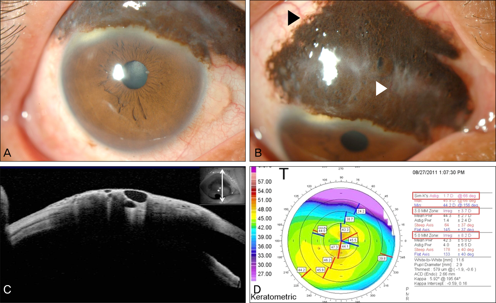
Figure 1. Preoperative photographs and examinations. (A, B) Photograph of conjunctival nevus. It sized 16 (horizontal) × 9 mm (vertical) with intralesional clear cysts, feeder vessels (black arrow head, ▶) and intrinsic vessels (white arrow head, ▷). (C) Anterior OCT (Optical Coherence Tomography) of conjunctival nevus. They show multiple clear cysts in the mass. (D) Preoperative topography of right eye. It shows 1.7 diopter of corneal astigmatism.

Figure 2. Postoperative photographs and examinations. (A) Two weeks after surgery. (B) Postoperative topography of right eye (two months after surgery). Corneal astigmatism was decreased to 0.7 diopter from 1.7 diopter. (C, D) Two months after surgery. No recurrence conjunctival nevus (D).
Reference
-
References
1. Shields CL, Fasiuddin AF, Mashayekhi A, Shields JA.Conjunctival nevi: clinical features and natural course in 410 consecutive patients. Arch Ophthalmol. 2004; 122:167–75.2. Shields CL, Regillo AC, Mellen PL. . Giant conjunctival ne-vus: clinical features and natural course in 32 cases. JAMA Ophthalmol. 2013; 131:857–63.3. Tomita M, Goto H, Muramatsu R, Usui M.Treatment of large con-junctival nevus by resection and reconstruction using amniotic membrane. Graefes Arch Clin Exp Ophthalmol. 2006; 244:761–4.
Article4. Shields CL, Demirci H, Karatza E, Shields JA.Clinical survey of 1643 melanocytic and nonmelanocytic conjunctival tumors. Ophthalmology. 2004; 111:1747–54.
Article5. Levecq L, De Potter P, Jamart J.Conjunctival nevi clinical features and therapeutic outcomes. Ophthalmology. 2010; 117:35–40.6. Shields CL, Markowitz JS, Belinsky I. . Conjunctival melano-ma: outcomes based on tumor origin in 382 consecutive cases. Ophthalmology. 2011; 118:389–95. e1-2.
Article7. Shields CL, Shields JA, Gündüz K. . Conjunctival melanoma: risk factors for recurrence, exenteration, metastasis, and death in 150 consecutive patients. Arch Ophthalmol. 2000; 118:1497–507.8. Oellers P, Karp CL.Management of pigmented conjunctival lesions. Ocul Surf. 2012; 10:251–63.9. Shields CL, Belinsky I, Romanelli-Gobbi M. . Anterior segment optical coherence tomography of conjunctival nevus. Ophthalmology. 2011; 118:915–9.
Article10. Jo DH, Lee MJ, Han YK, Kwon JW.Surgical treatment of ex-tensive conjunctival melanocytic nevus mimicking conjunctival melanoma. J Korean Ophthalmol Soc. 2010; 51:764–8.
Article
- Full Text Links
-
- Actions
-
Cited
- CITED
-
- Close
- Share
-
- Similar articles
-
- Malignant Melanoma Developed from Giant Congenital pigmented Nevus: Report of a Case
- Malignant Melanoma Arising from Giant Congenital Pigmented Nevus
- A Case of Malignant Melanoma in Children
- A Case of Malignant Melanoma Arising from Giant Congenital Melanocytic Nevus in a 4-year-old Girl
- A Conjunctival Malignant Melanoma in a Child
