A Case of Pleomorphic Adenoma Arising in the Ectopic Lacrimal Gland of the Lower Eyelid
- Affiliations
-
- 1Department of Ophthalmology, Chonnam National University Medical School, Gwangju, Korea. kcyoon@jnu.ac.kr
Abstract
- PURPOSE
To report a case of pleomorphic adenoma arising in the ectopic lacrimal gland of the lower eyelid.
CASE SUMMARY
A 36-year-old male presented with a gradually increasing mass in the left lower eyelid that had been growing for one year. A hard, non-tender, subcutaneous mass was palpated on the lateral one-third of the left lower eyelid, and there were no clinically specific signs. Orbital computed tomography demonstrated a well-demarcated 1 x 0.7 x 0.7-cm-sized mass with heterogeneous enhancement, and complete surgical resection was performed. The mass was non-adjacent to the conjunctiva and inferior tarsal plate. Histological examination showed glandular elements embedded in a myxoid stroma. The mass was diagnosed as pleomorphic adenoma arising in the ectopic lacrimal gland. At the postoperative 6-month follow-up, there was no recurrence or abnormal finding at the operation site.
CONCLUSIONS
Pleomorphic adenoma arising in the ectopic lacrimal gland should be considered as a differential diagnosis of eyelid masses.
MeSH Terms
Figure
-
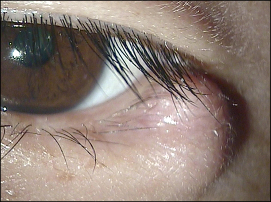
Figure 1. Clinical photograph of a 36-year-old man with subcutaneous mass in the left lateral lower eyelid.

Figure 2. Orbit CT shows a 1 × 0.7 × 0.7 cm-sized, round, well defined, heterogeneously enhancing mass. Axial (A) and coronal (B) views.
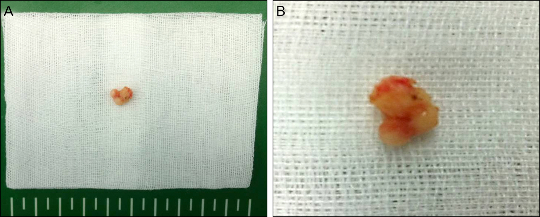
Figure 3. (A, B) A polypoid well encapsulated 1 × 0.7 × 0.5 cm-sized yellowish mass was completely excised.
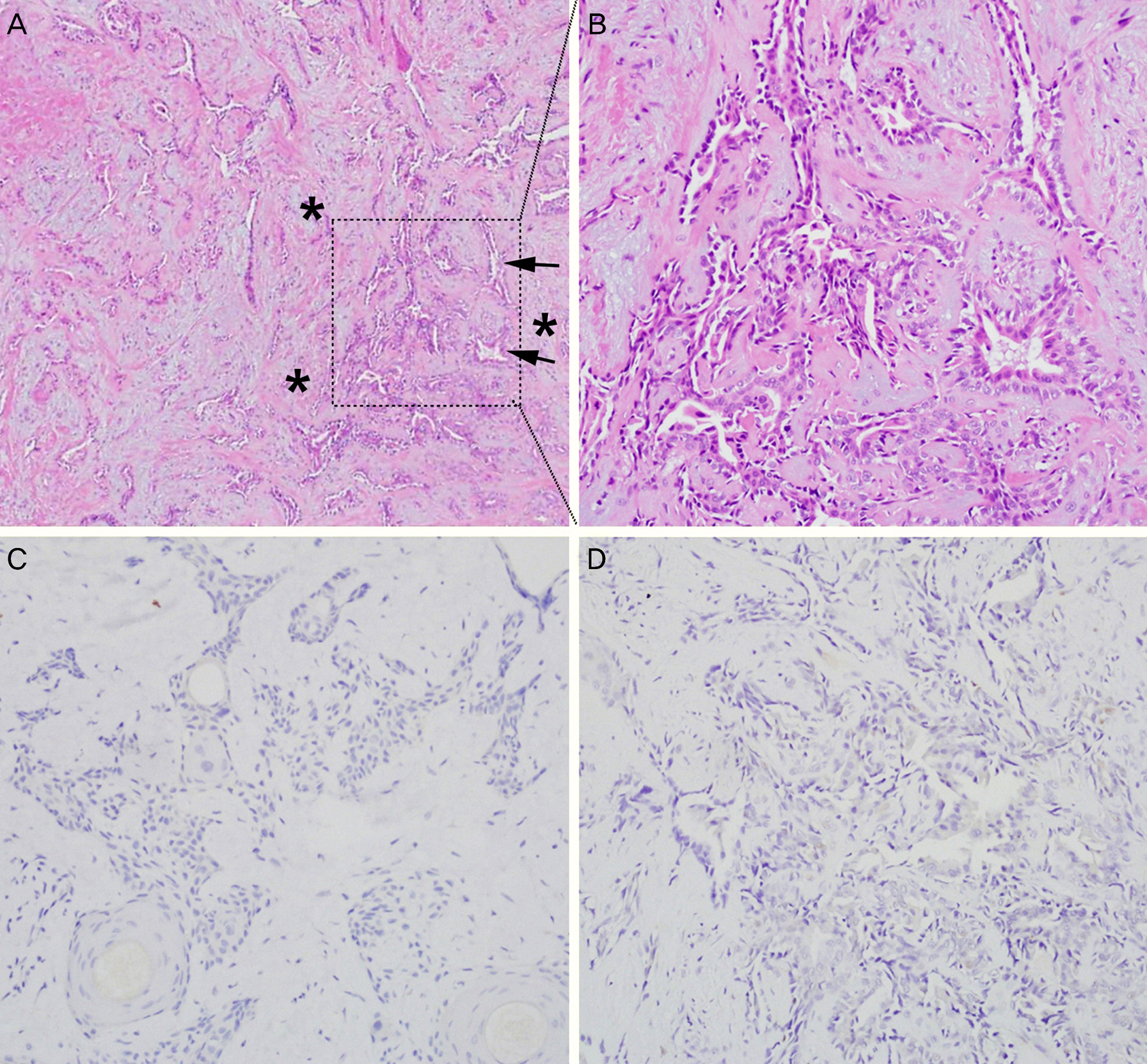
Figure 4. (A, B) Hematoxylin and eosin stained sections shows many glandular elements (arrows) embedded in myxoid ground substances (asterisks). (A) Hematoxylin and eosin stain, ×40. (B) Hematoxylin and eosin stain, ×200. (C) Tumor cells were not reacted with the antibodies against glial fibrillary acidic protein (immunostain, ×200). (D) Tumor cells were not reacted with the antibodies against S-100 protein (immunostain, ×200).
Reference
-
References
1. Shields JA, Blackwell B, Augsburber JJ, Flanagan JC. Classification and incidence of space-occupying lesions of the orbit. A survey of 645 biopsies. Arch Ophthalmol. 1984; 102:1606–11.2. Auran J, Jakobiec FA, Krebs W. Benign mixed tumor of the palpebral lobe of the lacrimal gland. Clinical diagnosis and appropriate surgical management. Ophthalmology. 1988; 95:90–9.3. Parks SL, Glover AT. Benign mixed tumors arising in the palpebral lobe of the lacrimal gland. Ophthalmology. 1990; 97:526–30.4. Jung BJ, Cho YK, La TY. A giant pleomorphic adenoma of lacrimal gland involving the palpebral lobe causing severe mechanical ptosis. J Korean Ophthalmol Soc. 2011; 52:241–5.
Article5. Tong JT, Flanagan JC, Eagle RC Jr, Mazzoli RA. Benign mixed tumor arising from an accessory lacrimal gland of Wolfring. Ophthal Plast Reconstr Surg. 1995; 11:136–8.
Article6. Venkataramayya K. Pleomorphic adenoma of Krause's gland. Indian J Ophthalmol. 1976; 23:38–9.7. Kapoor S, Sood GC, Kapoor MS, Aurora AL. Giant pleomorphic adenoma of accessory lacrimal gland. Indian J Ophthalmol. 1978; 25:52–3.8. Saini JS, Mukherjee AK, Naik P. Pleomorphic adenoma of Krause's gland in lower lid. Indian J Ophthalmol. 1985; 33:181–2.9. Patyal S, Banarji A, Bhadauria M, Gurunadh VS. Pleomorphic adenoma of a subconjunctival ectopic lacrimal gland. Indian J Ophthalmol. 2010; 58:245–7.
Article10. Shin KS, Kim YD, Lee HK. A case ofbenign mixed tumor presenting as a nodular eyelid lesion. J Korean Ophthalmol Soc. 1991; 32:101–5.11. Kim NJ, Choung HK, Khwarg SI. Benign mixed tumor arising from an accessory lacrimal gland in the inferior palpebral conjunctiva. J Korean Ophthalmol Soc. 2006; 47:1673–7.12. Rootman J, Mugent RA, Stewart B. Structure of the orbit: anatomic and imaging features. Rootman J, editor. Disease of the orbit. 2nd ed.Philadelphia: Lippincott Williams & Wilkins;2003. chap. 1.13. Ramlee N, Ramli N, Tajudin LS. Pleomorphic adenoma in the palpebral lobe of the lacrimal gland misdiagnosed as chalazion. Orbit. 2007; 26:137–9.
Article14. Nagao T, Sato E, Inoue R. . Immunohistochemical analysis of salivary gland tumors: application for surgical pathology practice. Acta Histochem Cytochem. 2012; 45:269–82.
Article15. Curran AE, Allen CM, Beck FM. . Distinctive pattern of glial fibrillary acidic protein immunoreactivity useful in distinguishing fragmented pleomorphic adenoma, canalicular adenoma and polymorphous low grade adenocarcinoma of minor salivary glands. Head Neck Pathol. 2007; 1:27–32.
Article16. Shah SS, Chandan VS, Wilbur DC, Khurana KK. Glial fibrillary acidic protein and CD57 immunolocalization in cell block preparations is a useful adjunct in the diagnosis of pleomorphic adenoma. Arch Pathol Lab Med. 2007; 131:1373–7.
Article17. Alyahya GA, Stenman G, Persson F. . Pleomorphic adenoma arising in an accessory lacrimal gland of Wolfring. Ophthalmology. 2006; 113:879–82.
Article18. Jung CK, Kim SM, Lee JY, Chung SK. Malignant change of pleomorphic adenoma. J Korean Ophthalmol Soc. 1997; 38:2251.19. Shields JA, Shields CL. Malignant transformation of presumed pleomorphic adenoma of lacrimal gland after 60 years. Arch Ophthalmol. 1987; 105:1403–5.
Article
- Full Text Links
-
- Actions
-
Cited
- CITED
-
- Close
- Share
-
- Similar articles
-
- Recurrent Pleomorphic Adenoma Arising from the Ectopic Lacrimal Gland in the Upper Eyelid
- Carcinoma expleomorphic adenoma of lacrimal gland
- An Ectopic Pleomorphic Adenoma in the Superficial Subcutaneous Layer of the Preauricular Area
- Ectopic pleomorphic adenoma on subcutaneous plane of the cheek
- Pleomorphic Adenoma of the Lacrimal Gland in a Child
