J Korean Ophthalmol Soc.
2011 Feb;52(2):157-162.
Long-Term Changes in Tilt, Decentration and Anterior Chamber Depth After Implantable Collamer Lens Insertion
- Affiliations
-
- 1Department of Ophthalmology and Visual Science, The Catholic University of Korea School of Medicine, Seoul, Korea. eyedoc@catholic.ac.kr
Abstract
- PURPOSE
To evaluate the stability of implantable collamer lens (ICL, Staar Surgical AG, Niau, Switzerland) by comparing changes of tilt, decentration and anteroir chamber depth after ICL implantation during 1 year.
METHODS
The results of 8 high myopic patients (16 eyes) that had received ICL implantation were retrospectively studied. Tilt and decentration of ICL were measured using an anterior eye segment analysis system (Scheimpflug camera, EAS-1000, Nidek, Japan). Anterior chamber depth was measured in both eyes preoperatively and postoperatively by Scheimpflug camera. The follow-up period was 1 year.
RESULTS
Tilt was 1.90 +/- 1.23degrees, 1.75 +/- 0.80degrees, 1.64 +/- 0.86degrees, 2.08 +/- 1.33degrees (p = 0.36) and decentration were 0.04 +/- 0.01 mm, 0.03 +/- 0.01 mm, 0.03 +/- 0.02 mm, 0.04 +/- 0.02 mm (p = 0.59) at 1 week, 1 month, 6 months and 1 year respectively. Tilt and decentration showed no significant change after ICL implantation. The average anterior chamber depth was 2.85 +/- 0.26 mm preoperatively, and 2.17 +/- 0.39 mm, 2.06 +/- 0.31 mm, 2.06 +/- 0.34 mm, 2.04 +/- 0.35 mm at 1 week, 1 month, 6 months and 1 year respectively. Anterior chamber depth became narrow after ICL implantation (p = 0.02), but showed no significant narrowing postoperatively (p = 0.08).
CONCLUSIONS
The IOL position remained stable, with no significant changes for an extended period of tilt, decentration, or anterior chamber depth after ICL implantation.
Figure
-

Figure 1. Measuring tilt and decentration of implantable collamer lens (ICL) based on Scheimpflug image.
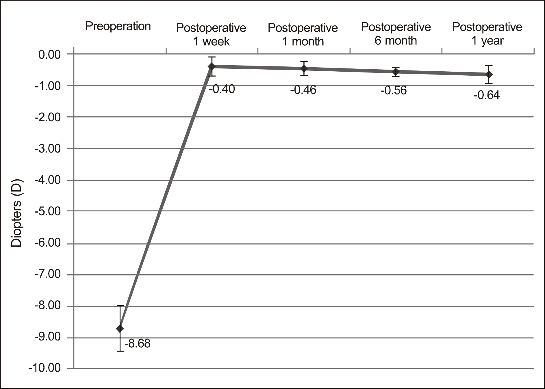
Figure 2. Changes in refractive power (spherical equivalent) after ICL implantation in patients with high myopia.
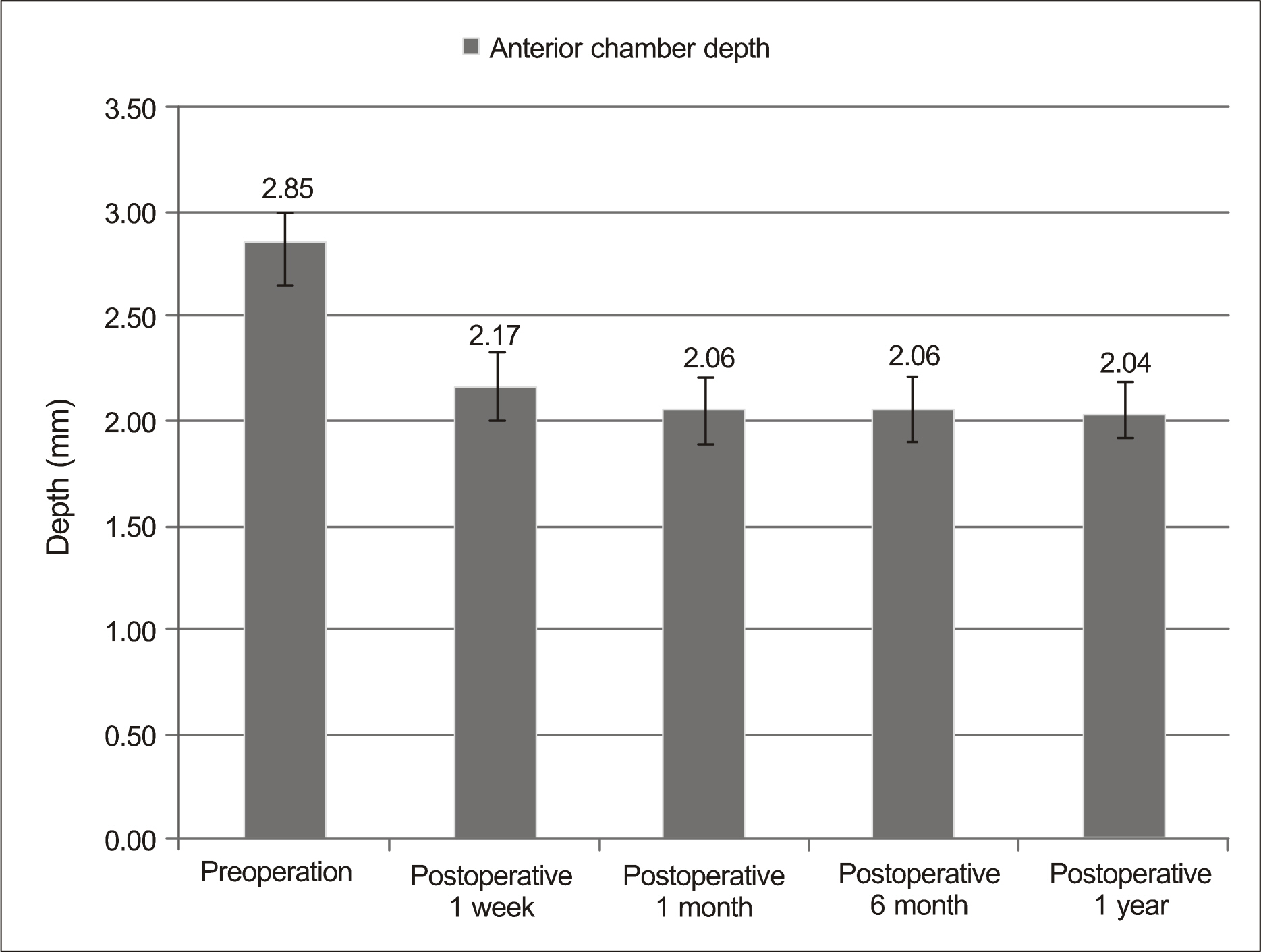
Figure 3. Changes in anterior chamber depth (mm) before and after ICL implantation in high myopic patients.
Reference
-
References
1. Marcos S. Aberrations and visual performance following standard laser vision correction. J Refract Surg. 2001; 17:S596–601.
Article2. Fyodorov SN, Zuyev VK, Aznabayev BM. Intraocular correction of high myopia with negative posterior chamber lens. Ophthal- mosurgery. 1991; 3:57–8.3. Sanders DR, Vukich JA; ICL in Treatment of Myopia (ITM) Study Group. Incidence of lens opacities and clinically significant cataracts with the implantable contact lens: comparison of two lens designs. J Refract Surg. 2002; 18:673–82.4. Gonvers M, Othenin-Girard P, Bornet C, Sickenberg M. Implantable contact lens for moderate to high myopia: short-term followup of 2 models. J Cataract Refract Surg. 2001; 27:380–8.5. Dejaco-Ruhswurm I, Scholz U, Pieh S, et al. Long-term endothelial changes in phakic eyes with posterior chamber intraocular lenses. J Cataract Refract Surg. 2002; 28:1589–93.
Article6. Edelhauser HF, Sanders DR, Azar R, et al. Corneal endothelial assessment after ICL implantation. J Cataract Refract Surg. 2004; 30:576–83.
Article7. Risco JM, Cameron JA. Dislocation of a phakic intraocular lens. Am J Ophthalmol. 1994; 118:666–7.
Article8. Ibrahim O, Waring GO 3rd. Successful exchange of dislocated phakic intraocular lens. J Refract Surg. 1995; 11:282–3.9. Alió JL, de la Hoz F, Pérez-Santonja JJ, et al. Phakic anterior chamber lenses for the correction of myopia: a 7-year cumulative analysis of complications in 263 cases. Ophthalmology. 1999; 106:458–66.10. Budo C, Hessloehl JC, Izak M, et al. Multicenter study of the Artisan phakic intraocular lens. J Cataract Refract Surg. 2000; 26:1163–71.
Article11. Pérez-Santonja JJ, Bueno JL, Zato MA. Surgical correction of high myopia in phakic eyes with Worst-Fechner myopia intraocular lenses. J Refract Surg. 1997; 13:268–81.
Article12. Wiechens B, Winter M, Haigis W, et al. Bilateral cataract after phakic posterior chamber top hat-style silicone intraocular lens. J Refract Surg. 1997; 13:392–7.
Article13. Khan AJ, Percival SP. 12 year results of a prospective trial comparing poly(methyl methacrylate) and poly (hydroxyethyl methacrylate) intraocular lenses. J Cataract Refract Surg. 1999; 25:1404–7.14. Rosen E, Gore C. Staar Collamer posterior chamber phakic intraocular lens to correct myopia and hyperopia. J Cataract Refract Surg. 1998; 24:596–606.
Article15. Zaldivar R, Davidorf JM, Oscherow S. Posterior chamber phakic intraocular lens for myopia of −8 to −19 diopters. J Refract Surg. 1998; 14:294–305.
Article16. Zaldivar R, Oscherow S, Ricur G. The STAAR posterior chamber phakic intraocular lens. Int Ophthalmol Clin. 2000; 40:237–44.
Article17. Sanders DR, Brown DC, Martin RG, et al. Implantable contact lens for moderate to high myopia: phase 1 FDA clinical study with 6 months followup. J Cataract Refract Surg. 1998; 24:607–11.18. Sarver EJ, Sanders DR, Vukich JA. Image quality in myopic eyes corrected with laser in situ keratomileusis and phakic intraocular lens. J Refract Surg. 2003; 19:397–404.
Article19. Sanders DR, Vukich JA, Doney K, Gaston M. U.S. Food and Drug Administration clinical trial of the implantable contact lens for moderate to high myopia. Ophthalmology. 2003; 110:255–66.
Article20. Uusitalo RJ, Aine E, Sen NH, Laatikainen L. Implantable contact lens for high myopia. J Cataract Refract Surg. 2002; 28:29–36.
Article21. Han SY, Lee KH. Long term effect of ICL implantation to treat high myopia. J Korean Ophthalmol Soc. 2007; 48:465–72.22. Gonvers M, Bornet C, Othenin-Girard P. Implantable contact lens for moderate to high myopia: relationship of vaulting to cataract formation. J Cataract Refract Surg. 2003; 29:918–24.23. Kwon SM, Oh HC, Lee DJ, et al. Comparison of anterior segment parameters in angle-closure glaucoma using Scheimpflug camera. J Korean Ophthalmol Soc. 2009; 50:128–34.24. Catalano RA, Kassoff A. Irreversible intraocular lens vaulting with pupillary-block glaucoma. Am J Ophthalmol. 1986; 101:735–6.
Article25. Kamiya K, Shimizu K, Komatsu M. Factors affecting vaulting after implantable collamer lens implantation. J Refract Surg. 2009; 25:259–64.
Article26. Kojima T, Maeda M, Yoshida Y, et al. Posterior chamber phakic implantable collamer lens: changes in vault during 1 year. J Refract Surg. 2010; 26:327–32.
Article27. Kamiya K, Shimizu K, Kawamorita T. Changes in vaulting and the effect on refraction after phakic posterior chamber intraocular lens implantation. J Cataract Refract Surg. 2009; 35:1582–6.
Article28. Chun YS, Lee JH, Lee JM, Park IK. IOP and gonioscopic changes after implantable contract lens implantation in myopic eyes. J Korean Ophthalmol Soc. 2005; 46:336–44.29. Sasaki K, Sakamoto Y, Shibata T, et al. Measurement of postoperative intraocular lens tilting and decentration using Scheimpflug images. J Cataract Refract Surg. 1989; 15:454–7.
Article30. Baumeister M, Neidhardt B, Strobel J, Kohnen T. Tilt and decentration of three-piece foldable high-refractive silicone and hydrophobic acrylic intraocular lenses with 6-mm optics in an intra-individual comparison. Am J Ophthalmol. 2005; 140:1051–8.
Article
- Full Text Links
-
- Actions
-
Cited
- CITED
-
- Close
- Share
-
- Similar articles
-
- Decentration, Tilt and Anterior Chamber Depth: Aspheric vs Spheric Acrylic Intraocular Lens
- Diurnal Variation in the Depth of the Anterior Chamber and Correlation between the Depth of the Anterior Chamber and Intraocular Pressure
- The Relationship of the Lens Density with the Lens Thickness and the Anterior Chamber Depth
- Comparison of the Stability Between Three-piece and Single-piece Aspheric Intraocular Lenses
- Comparison of Decentration and Tilt of Sensar(R) with Acrysof
