J Korean Ophthalmol Soc.
2010 Mar;51(3):463-468.
Bilateral Serous Retinal Detachment Associated With Alport's Syndrome
- Affiliations
-
- 1Department of Ophthalmology, Kangdong Sacred Heart Hospital, Hallym University College of Medicine, Seoul, Korea. sungpyo@hananet.net
Abstract
- PURPOSE
To report a case of bilateral serous retinal detachment associated with Alport's syndrome that resolved following intensive blood pressure control and electrolyte imbalance correction.
CASE SUMMARY
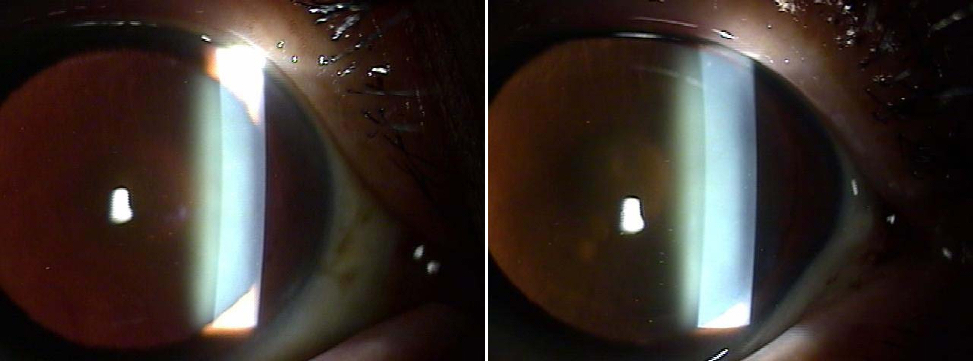
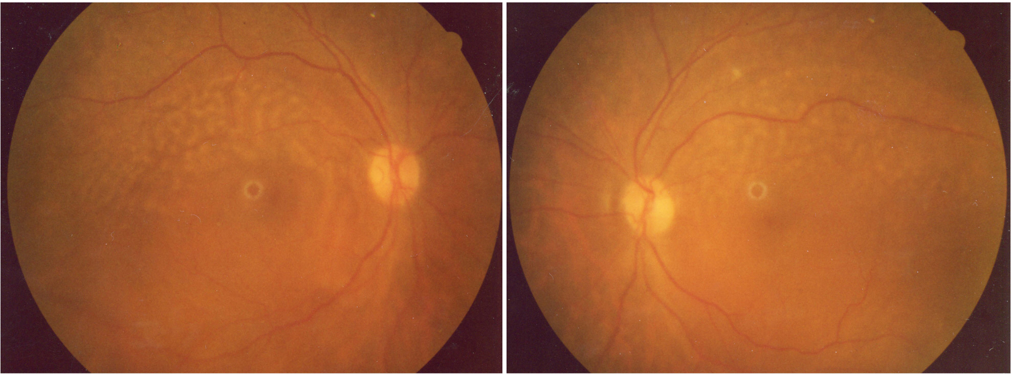
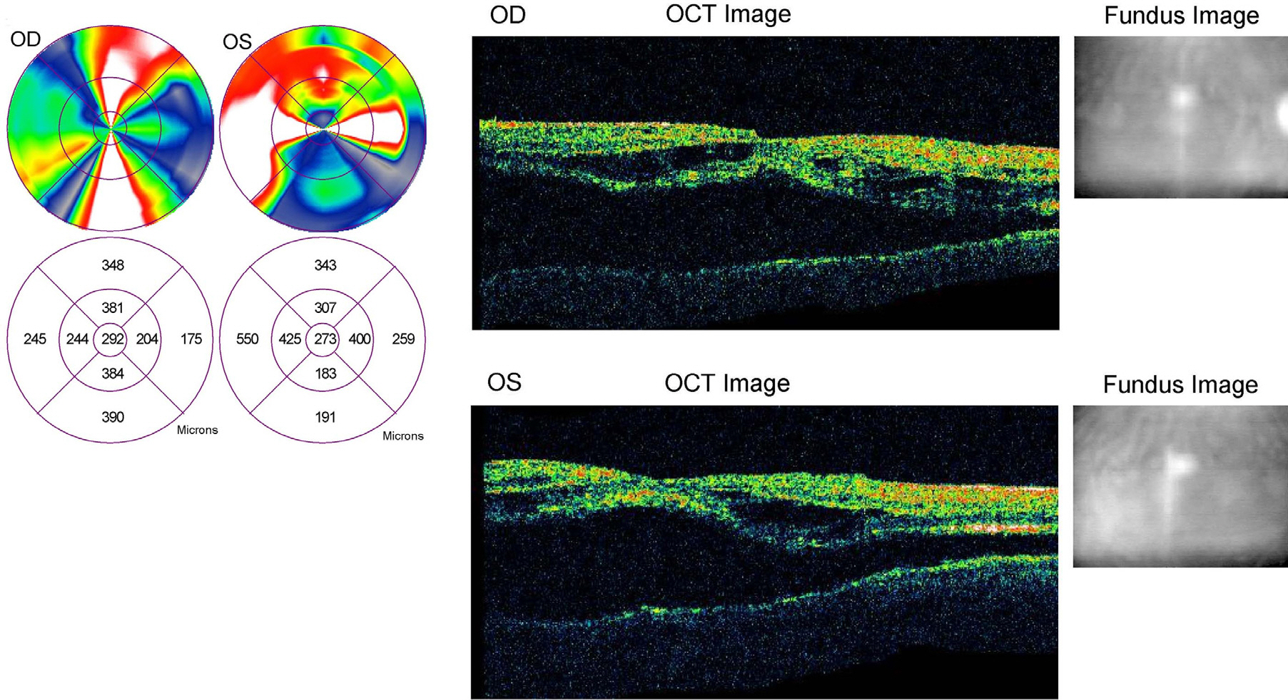
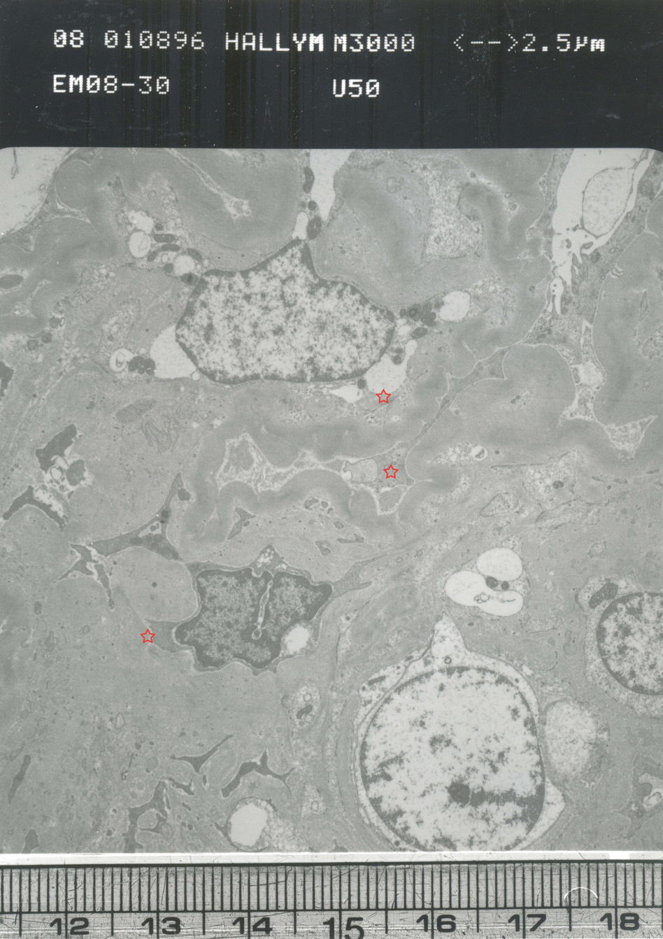
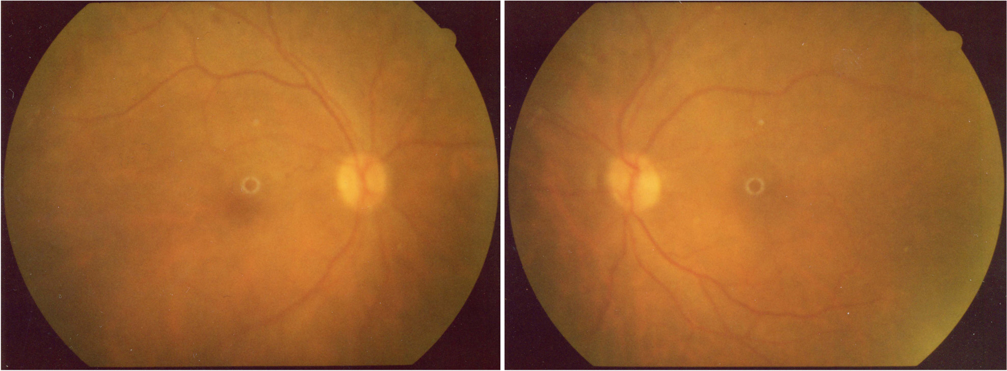
A 50-year-old male patient presented with bilateral lenticonus and bilateral serous retinal detachment. Bilateral serous retinal detachment with retinal flecks characteristic of Alport's syndrome appeared along with the development of chronic renal failure and hypertension. The following kidney biopsy revealed Alport's syndrome. After 14 days, the serous detachment resolved and vision recovered following intensive blood pressure control and electrolyte imbalance correction fundus and FA results were nearly normal.
CONCLUSIONS
In this case, bilateral serous retinal detachment in Alport's syndrome resolved with intensive blood pressure control and electrolyte imbalance correction. To the author's knowledge, this is the first case in South Korea with documentation of the onset and resolution of bilateral serous retinal detachment in Alport's syndrome.
MeSH Terms
Figure
Reference
-
References
1. Alport AC. Hereditary familial congenital hemorrhagic nephritis. Brit Med J. 1927; 1:504.2. Gass JD. Stereoscopic Atlas of Macular Diseases, Diagnosis and Treatment. 4th ed.St Louis: Mosby Co.;1997. p. 303.3. Duke-Elder S. System of Ophthalmology. St. Louis: Mosby Co.;1969. p. 696.4. Kenjiro Y, Soh F, Motohiro K, et al. Bilateral Serous Retinal Detachment Associated with Alport's Syndrome. Ophthalmologica. 2000; 214:301–4.
Article5. Yoshikawa N, White RH, Cameron AH. Familial hematuria: clinicopathological correlations. Clin Nephrol. 1982; 17:172–82.6. Mayers JC, Jones TA, Pohjolainen ER, et al. Molecular cloning of α5(IV) collagen and assignment of the gene to the region of the X chromosome containing the Alport syndrome locus. Am J Hum Genet. 1990; 46:1024–33.7. Hostikka SL, Eddy RL, Byers MG, et al. Identification of a distinct type IV collagen alpha chain with restricted kidney distribution and assignment of its gene to the locus of X chromosome-linked Alport syndrome. Proc Natl Acad Sci USA. 1990; 87:1606–10.
Article8. Gubler M, Levy M, Brover M, et al. Alport's syndrome, A report of 58 cases and a review of the literature. Am J Med. 1981; 70:493.9. Singh DS, Bisht DB, Kapoor S, et al. Lenticonus in Alport's syndrome, A family study. Acta Ophthalmol. 1977; 55:164.10. Choi J, Na K, Bae S, Roh G. Anterior lens capsule abnormalities in Alport syndrome. Korean J Ophthalmol. 2005; 19:84–9.
Article11. Johnstone WW. Anterior lenticonus. Am J Ophthalmol. 1963; 56:991.
Article12. Gehrs KM, Pollock SC, Zilkha G. Clinical features and abdominal of Alport retinopathy. Retina. 1995; 15:305–11.13. Usui T, Ichibe M, Hasegawa S, et al. Symmetrical reduced retinal thickness in a patient with Alport syndrome. Retina. 2004; 24:977–9.
Article14. Inomata H, Oka Y. Ultrastructural alterations of retinal blood vessels in renal retinopathy, Two cases who had undergone and not undergone hemodialysis, and pathogenesis for metastatic abdominal in the vessel walls. Nippon Ganka Gakkai Zasshi. 1972; 76:1079–88.
- Full Text Links
- Actions
-
Cited
- CITED
-
- Close
- Share
- Similar articles
-
- Spontaneous Resolution of Post-Traumatic Bilateral Serous Retinal Detachment in Childrens
- A case of Atypical Central Serous Chorioretinopathy with Bullous Retinal Detachment
- Laser Photocoaculation Treatment in a Case of Circumscribged Choroidal hmangioma Associated with Serous Retinal Detachment
- Takayasu's Arteritis Associated with Serous Retinal Detachment
- A Case of Central Serous Chorioretinopathy with Bullous Retinal Detachment

