J Korean Ophthalmol Soc.
2010 Mar;51(3):340-346.
Analysis of Postoperative Macular Edema in Cataract Patients with Diabetes using Optical Coherence Tomography
- Affiliations
-
- 1Department of Ophthalmology and Visual Science, The Catholic University of Korea College of Medicine, Seoul, Korea. eyedoc@catholic.ac.kr
Abstract
- PURPOSE
To evaluate the incidence and progression of macular edema (ME) and associated risk factors in diabetic patients.
METHODS
In a prospective study, 66 eyes were assessed by optical coherence tomography (OCT) and best-corrected visual acuity was checked before operation at one and three months after operation. ME was defined as an increase of central macular thickness (CMT) by 30% or more after surgery than before operation, as measured by OCT.
RESULTS
The incidence of ME in diabetic patients was 8.8%. The increment of CMT at three months after cataract surgery was statistically significant in the patients of diabetic duration> or =10 years (p=0.049). But insulin treatment, the severity of diabetic retinopathy, diabetic nephropathy and hemoglobin A1C were not significant risk factors for ME.
CONCLUSIONS
The OCT might be useful to assess the ME after cataract surgery in diabetic patients. In the patients who had long been suffered from diabetes, the incidence of ME could be higher, so cataract surgery should be carefully considered.
MeSH Terms
Figure
-
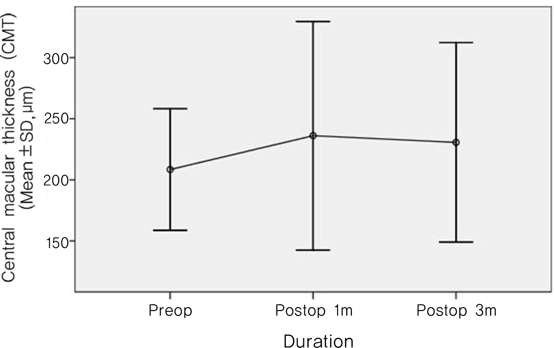
Figure 1. Mean central macular thickness of study eyes at 1 and 3 months preoperatively after cataract surgery.
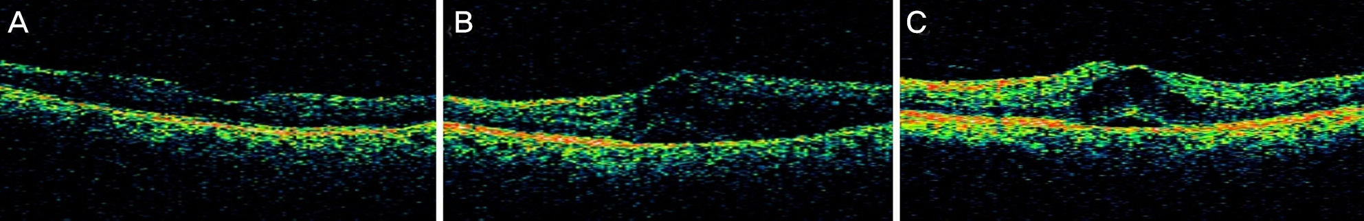
Figure 2. Representative range of cystoids abnormalities of study eyes with macular edema. (A) Preoperative optical coherence tomography (OCT). (B) Post-operative 1 month OCT. (C) Post-operative 3 months OCT.
Reference
-
References
1. Dowler JG, Hykin PG, Lightman SL, Hamilton AM. Visual abdominal following extracapsular cataract extraction in diabetes: a metaanalysis. Eye. 1995; 9:313–7.2. Zaczek A, Olivestedt G, Zetterstrom C. Visual outcome after abdominal and IOL implantation in diabetic patients. Br J Ophthalmol. 1999; 83:1036–41.3. Cunliffe IA, Flanagan DW, George ND, et al. Extracapsular abdominal surgery with lens implantation in diabetics with and without proliferative retinopathy. Br J Ophthalmol. 1991; 75:9–12.4. Pollack A, Dotan S, Oliver M. Progression of diabetic retinopathy after cataract extraction. Br J Ophthalmol. 1991; 75:547–51.
Article5. Henricsson M, Heijl A, Janzon L. Diabetic retinopathy before and after cataract surgery. Br J Ophthalmol. 1996; 80:789–93.
Article6. Kato S, Fukada Y, Hori S, et al. Influence of phacoemulsification and intraocular lens implantation on the course of diabetic retinopathy. J Cataract Refract Surg. 1999; 25:788–93.
Article7. Johnson RN, Howard SH, McDonald R, et al. Fluorescein abdominalgraphy: basic principles and interpretation. Ryan SJ, editor. Retina. revised ed.St. Louis: Mosby;2001. 2:chap. 55.8. Flesner P, Sander B, Henning V, et al. Cataract surgery on diabetic patients: a prospective evaluation of risk factors and complications. Acta Ophthalmol Scand. 2002; 80:19–24.
Article9. Hayashi K, Igarashi C, Hirata A, et al. Changes in diabetic abdominal oedema after phacoemulsification surgery. Eye. 2009; 23:389–96.10. Kim SJ, Equi R, Bressler NM. Analysis of macular edema after abdominal surgery in patients with diabetes using optical coherence tomography. Ophthalmology. 2007; 114:881–89.11. Early Treatment Diabetic Retinopathy Study Research Group. Photocoagulation for diabetic macular edema: Early Treatment Diabetic Retinopathy Study report number 1. Arch Ophthalmol. 1985; 103:1796–806.12. Hee MR, Puliafito CA, Wong C, et al. Quantitative assessmentof macular edema with optical coherence tomography. Arch Ophthalmol. 1995; 113:1019–29.13. Goebel W, Kretzchmar-Gross T. Retinal thickness in diabetic abdominal: a study using optical coherence tomography (OCT). Retina. 2002; 22:759–67.14. Chen CH, Liu YC, Wu PC. The combination of intravitreal abdominal and phacoemulsification surgery in patients with abdominal and coexisting diabetic macular edema. J Ocul Pharmacol Ther. 2009; 25:83–9.15. Cardillo J, Melo LA Jr, Costa RA, et al. Comparison of abdominal versus posterior abdominal's capsule injection of triamcinolone acetonide for diffuse diabetic macular edema. Ophthalmology. 2005; 112:1557–63.16. Larsson J, Zhu M, Sutter F, Gillies MC. Relation between reduction of foveal thickness and visual acuity in diabetic macular abdominal treated with intravitreal triamcinolone. Am J Ophthalmol. 2005; 139:802–6.17. Nelson ML, Martidis A. Managing cystoid macular edema after cataract surgery. Curr Opin Ophthalmol. 2003; 14:39–43.
Article18. Ray S, D'Amico DJ. Pseudophakic cystoid macular edema. Semin Ophthalmol. 2002; 17:167–80.
Article
- Full Text Links
-
- Actions
-
Cited
- CITED
-
- Close
- Share
-
- Similar articles
-
- Changes in Macular Thickness after Cataract Surgery According to Optical Coherence Tomography
- Analysis of Vitrectomy Results in Diabetic Macular Edema using Optical Coherence Tomography
- Evaluation of Changes of Macular Thickness in Diabetic Retinopathy after Cataract Surgery
- A Case of Secondary Macular Hole Formation after Phacoemulsification in a Vitrectomized Eye
- Risk Factors for Diffuse Diabetic Macular Edema as Classified by Optical Coherence Tomography
