J Korean Ophthalmol Soc.
2008 Nov;49(11):1829-1838.
Ophthalmic Manifestations in Patients With Neurofibromatosis
- Affiliations
-
- 1Department of Ophthalmology, Ajou University School of Medicine, Suwon, Korea. yhchang@ajou.ac.kr
Abstract
- PURPOSE
To report the ophthalmic manifestations of neurofibromatosis in Korea.
METHODS
Ophthalmologic examinations were performed from November 2001 to January 2008 for 153 consecutive patients who were diagnosed with neurofibromatosis according to the diagnostic criteria for neurofibromatosis. A retrospective analysis was performed according to the medical records of these 153 patients.
RESULTS
Seventy seven out of the 153 patients were men, 76 were women and the mean age was 20.44 +/- 14.34 years old. One hundred twelve were neurofibromatosis type 1 and six were neurofibromatosis type 2. Remained thirty five were segmental neurofibromatosis type 1. Ophthalmic manifestations of the neurofibromatosis type 1 were Lisch nodule (52.68%), high myopia (14.29%), plexiform neurofibroma in the orbit (4.46%), cafe au lait spots (4.46%) and optic glioma (3.58%). In the neurofibromatosis type 2, epiretinal membrane (33.33%) showed highest incidence and posterior subcapsular opacity (16.67%), Lisch nodule (16.67%), optic disc edema (16.67%), and optic nerve glioma (16.67%) were also noted. Lisch nodule (25.71%) was the most common ophthalmic finding in segmental neurofibromatosis type 1.
CONCLUSIONS
Lisch nodule, which was the most common manifestation of the neurofibromatosis type 1, was less manifested in our cases compared to the previous reports of western countries. In the neurofibromatosis type 2, epiretinal membrane and posterior subcapsular cataract showed higher incidence than those of other types of neurofibromatosis.
MeSH Terms
Figure
-
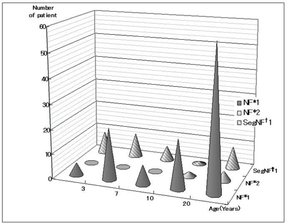
Figure 1. Distribution of age in neurofibromatosis;* Neurofibromatosis† Segmental neurofibromatosis.
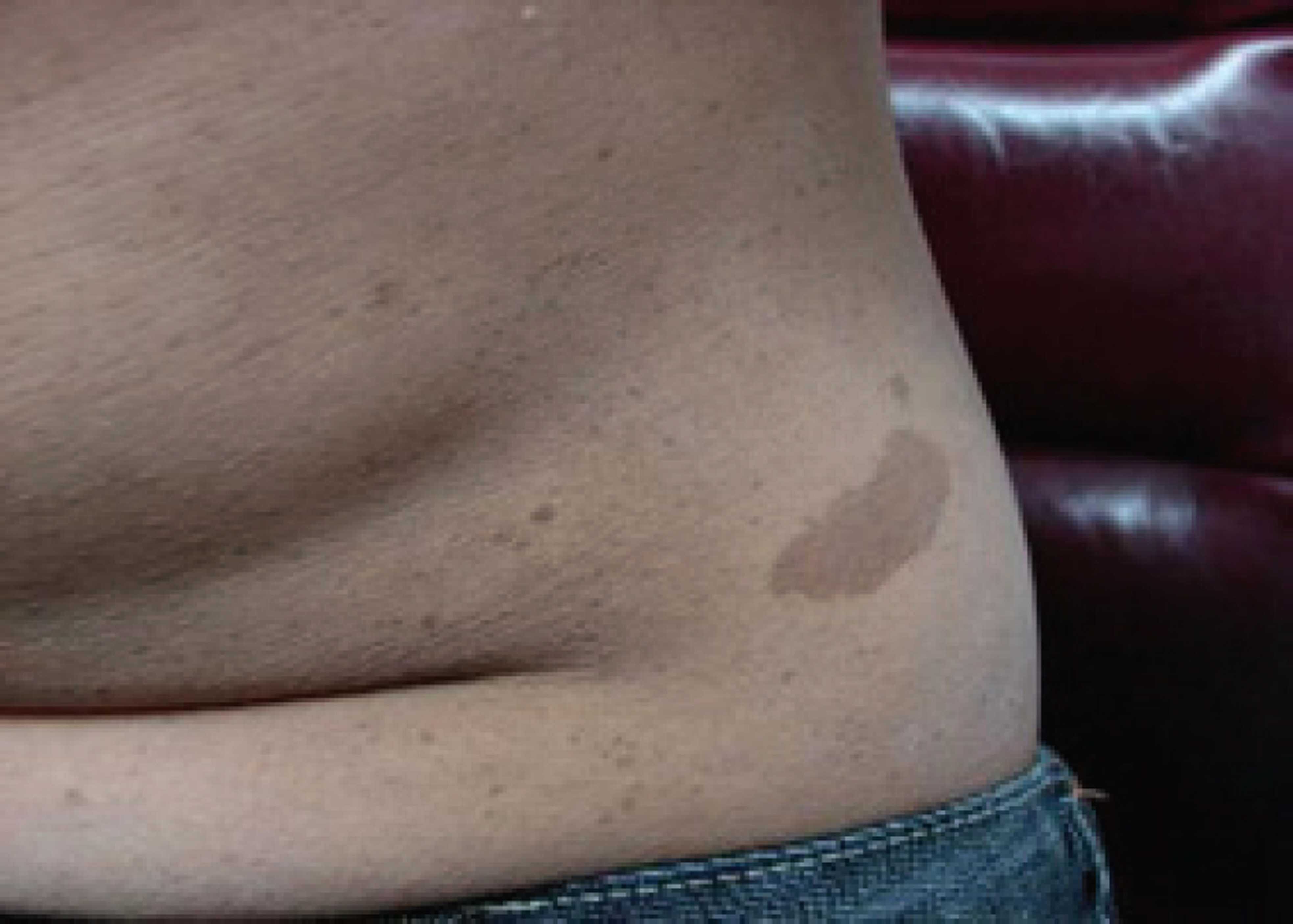
Firure 2. The photograph shows a large café au lait spots and freckles.
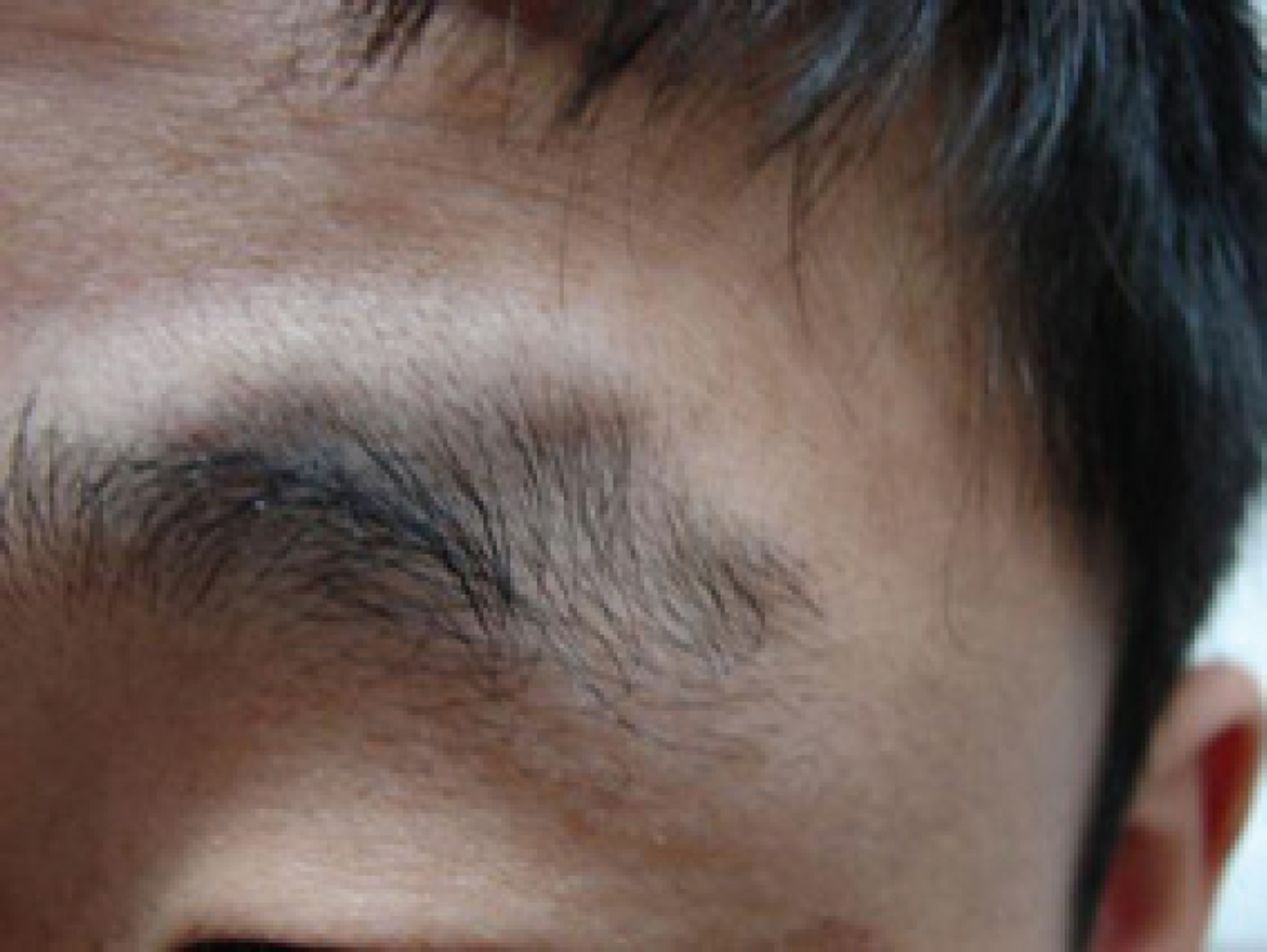
Figure 3. The photograph shows periorbital subcutaneous neurofibromas.
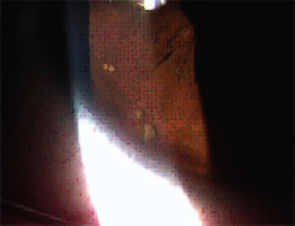
Figure 4. Anterior segment photograph shows multiple Lisch nodules on iris.

Figure 5. Unilateral optic glioma (arrow) in an individual affected by neurofibromatosis 1.
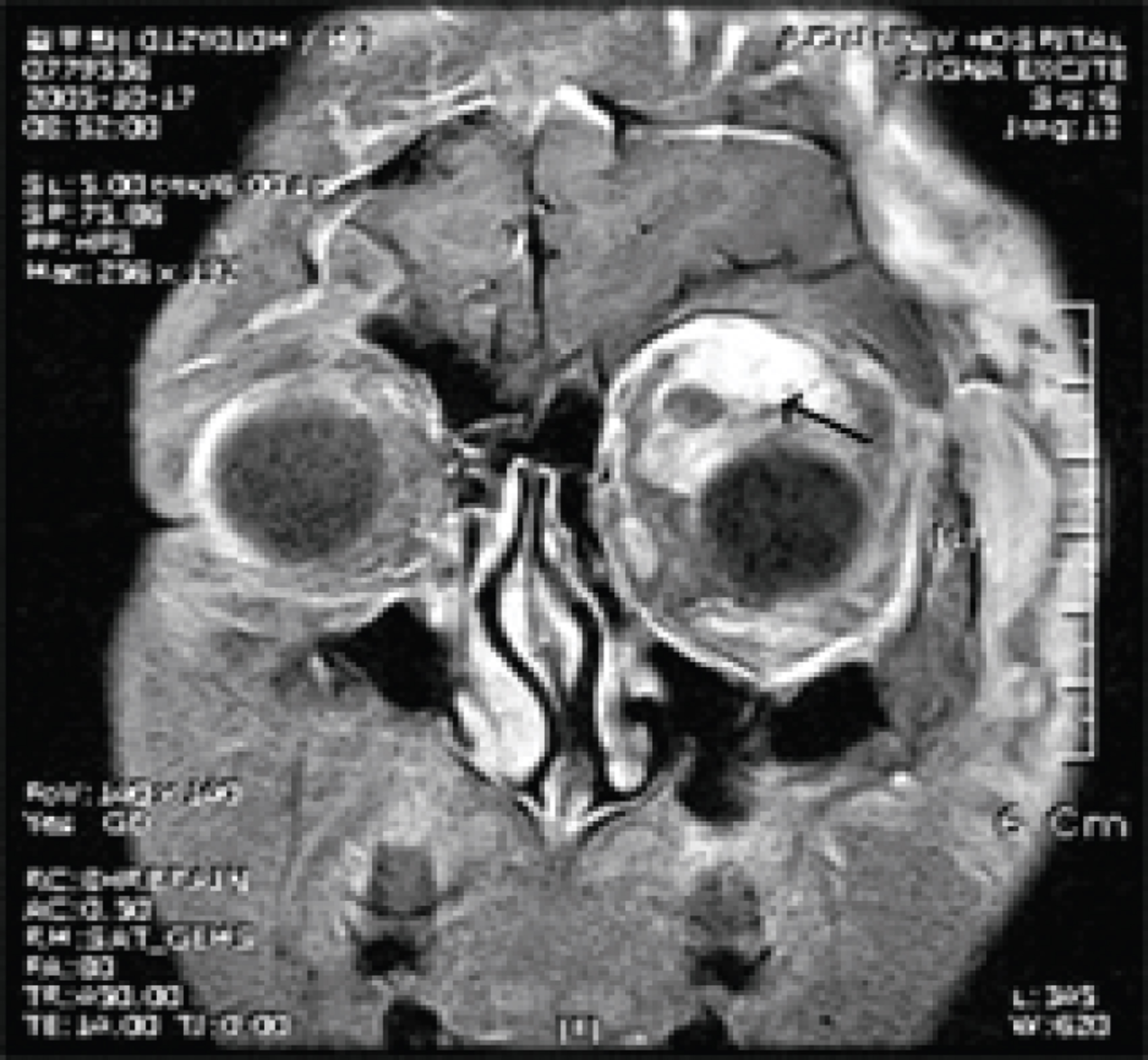
Figure 6. The MRI shows a plexiform neurofibroma (arrow) of the left orbit.

Figure 7. The photograph shows a elevated conjunctival neurofibroma.
Reference
-
References
1. Ahn HS. Hong CU. Pediatrics, 8th ed. Seoul: Daehan Printing & Publishing Co;2004; 1040–4.2. Menkes JH, Sernat HB. Child Neurology, 6th ed. Philadelphia: Lippincott Williams&Wilkins;2000; 859–84.3. Behrman RE, Kliegman RM, Jenson HB. Nelson textbook of Pediatrics, 17th ed. Philadelphia: Saunders;2004; 2015–9.4. Riccardi VM. Neurofibromatosis: Clinical heterogeneity. Curr Prob Cancer. 1982; 7:1–34.
Article5. Ropper AH, Brown RH. Adams and Victor’s principles of neurology, 8th ed. New York: McGraw-Hill;2005; 868–71.6. Saver S, Cestari DM. Neurofibromatosis type I: Genetics and clinical manifestations. Semin Ophthalmol. 2008; 23:45–51.7. Kreusel KM. Ophthamological manifestation in VKL and NF I: pathological and diagnostic implications. Fam Cancer. 2005; 4:43–7.8. National Institute of Health Consensus Development Conference. Neurofibromatosis: conference statement. Arch Neurol. 1988; 45:575–8.9. Baser ME, Friedman JM, Wallace AJ, et al. Evaluation of clinical diagnostic criteria for neurofibromatosis 2. Neurology. 2002; 59:1759–65.
Article10. Ruggieri M, Pavone P, Polizzi A, et al. Ophthalmological manifestations in segmental neurofibromatosis type1. Br J Ophthalmol. 2004; 88:1429–33.11. Listernick R, Mancini AJ, Charrow J. Segmental neurofibro matosis in childhood. Am J Med Genet A. 2003; 121:132–5.12. Gaonker CH, Mukherjee AK, Pokle M. Involvement of the eye and orbit in neurofibromatosis. Indian J Ophthalmol. 1992; 40:2–4.13. Huson S, Jones D, Beck L. Ophthalmic manifestations of neurofibromatosis. Br J Ophthalmol. 1987; 71:235–8.
Article14. Bosch MM, Boltshauser E, Harpes P, Landau K. Ophthalmo logic findings and long-term cource in patients with neurofibro matosis type 2. Am J Ophthalmol. 2006; 141:1068–77.15. Kim YH, Cho BC, Ko HK. 2 Cases of Neurofibromatosis. J Korean Ophthalmol Soc. 1982; 23:859–65.16. Kwon IT, Ohn YH, Shin HH. Retinal Finding of a Case of Neurofibromatosis. J Korean Ophthalmol Soc. 1994; 35:210–4.17. Yoon SW, Ahn BC. A Case of Orbital Neurilemoma Associated with Neurofibroma tosis. J Korean Ophthalmol Soc. 1999; 40:1993–7.18. Jung JW, Lee JH, Shyn KH, Chi MJ. A Case of Plexiform Neurofibroma with Severe Ptosis and Proptosis. J Korean Ophthalmol Soc. 2007; 48:725–30.19. Lee KH, Park JW, Yoon KC. A Case of Neurofibromatosis of the Orbit and Ocular Surface. J Korean Ophthalmol Soc. 2007; 48:1276–80.
Article20. Ferner RE. Neurofibromatosis 1 and neurofibromatosis 2: a twenty first century perspective. Lancet Neurol. 2007; 6:340–51.
Article21. Richetta A, Giustini S, Recupero SM, et al. Lisch nodules of the iris in neurofibromatosis type 1. J Eur Acad Dermatol Venereol. 2004; 18:342–4.
Article22. Albert DM, Green WR, Zimbric ML, et al. Iris melanocyte numbers in Asian, African-American, and Caucacian irides. Trans Am Ophthalmol Soc. 2003; 101:217–22.23. Arigon V, Binaghi M, Sabouret C, et al. Usefulness of systematic ophthalmologic investigations in neurofibromatosis 1: a cross sectional study of 211 patients. Eur J Ophthalmol. 2002; 12:413–8.24. Huson S, Jones D, Beck L. Ophthalmic manifestations of neurofibromatosis. Br J Ophthalmol. 1987; 71:235–8.
Article25. Payne MS, Nadell JM, Lacassie Y, Tilton AH. Congenital glaucoma and neurofibromatosis in a monozygotic twin: case report and review of the literature. J Child Neurolol. 2003; 18:504–8.
Article26. Rosser T, Packer RJ. Intracranial neoplasms in children with neurofibromatosis 1. J Child Neurol. 2002; 17:630–7.27. Ruggieri M, Huson SM. The clinical and diagnostic implications of mosaicism in the neurofibromatoses. Neurology. 2001; 56:1434–43.28. Uhrin SR. Segmental neurofibromatosis. South Med J. 1980; 73:526–7.29. Tinschert S, Naumann I, Stegmann E, et al. Segmental neurofibromatosis is caused by somatic mutation of the neurofibromatosis type 1 (NF1) gene. Eur J Hum Genet. 2000; 8:455–9.30. Radtke HB, Sebold CD, Allison C, et al. Neurofibromatosis type 1 in genetic counseling practice: recommendations of the National Society of Genetic Counselors. J Genet Couns. 2007; 16:387–407.
Article31. Messiaen LM, Callens T, Mortier G, et al. Exhaustive mutation analysis of the NF1 gene allows identification of 95% of mutations and reveals a high frequency of unusual splicing defects. Hum Mutat. 2000; 15:541–55.
- Full Text Links
-
- Actions
-
Cited
- CITED
-
- Close
- Share
-
- Similar articles
-
- A Case of Neurofibromatosis Associated with Moyamoya Disease
- Pseudo-Mascularization of the Phallus: The Clitoral Involvement of von Recklinghausen`s Neurofibromatosis
- A Case of Eccrine Spiradenoma in a Patient with Neurofibromatosis
- Two Cases of Neurofibrosarcoma Arising from Cutaneous Neurofibroma in Patients with Neurofibromatosis
- The radiologic findings of neurofibromatosis
