Use of Autologous Tenon's Capsule Graft for Repair of Traumatic Scleral Perforation
- Affiliations
-
- 1Department of Ophthalmology, Gangnam Sacred Hospital, College of Medicine, Hallym University, Seoul, Korea. eyedrnam@naver.com
Abstract
- PURPOSE
To report the use of autologous Tenon's capsule graft for repair of scleral defects caused by traumatic scleral perforation.
CASE SUMMARY
An 81-year-old man presented with loss of vision in his right eye after a perforating injury caused by a cow horn. Examination showed a laceration of the sclera at 12 o'clock approximately 5~6 mm in length, and a uveal tissue was prolapsed into the wound. The best corrected visual acuity was 0.1. Primary repair of the eye was insufficient because of tissue loss. The inferonasal Tenon's capsule graft was carefully dissected from the sclera and tailored to fit the defect. The graft was covered with a conjunctival flap. The scleral defect was successfully closed with the autologous Tenon's capsule graft. Three months after grafting, phacoemulsification with intraocular lens implantation was performed.
CONCLUSIONS
Autologous Tenon's capsule graft is an effective measure to repair traumatic scleral defects and is useful when patch grafts are unexpectedly needed.
MeSH Terms
Figure
-
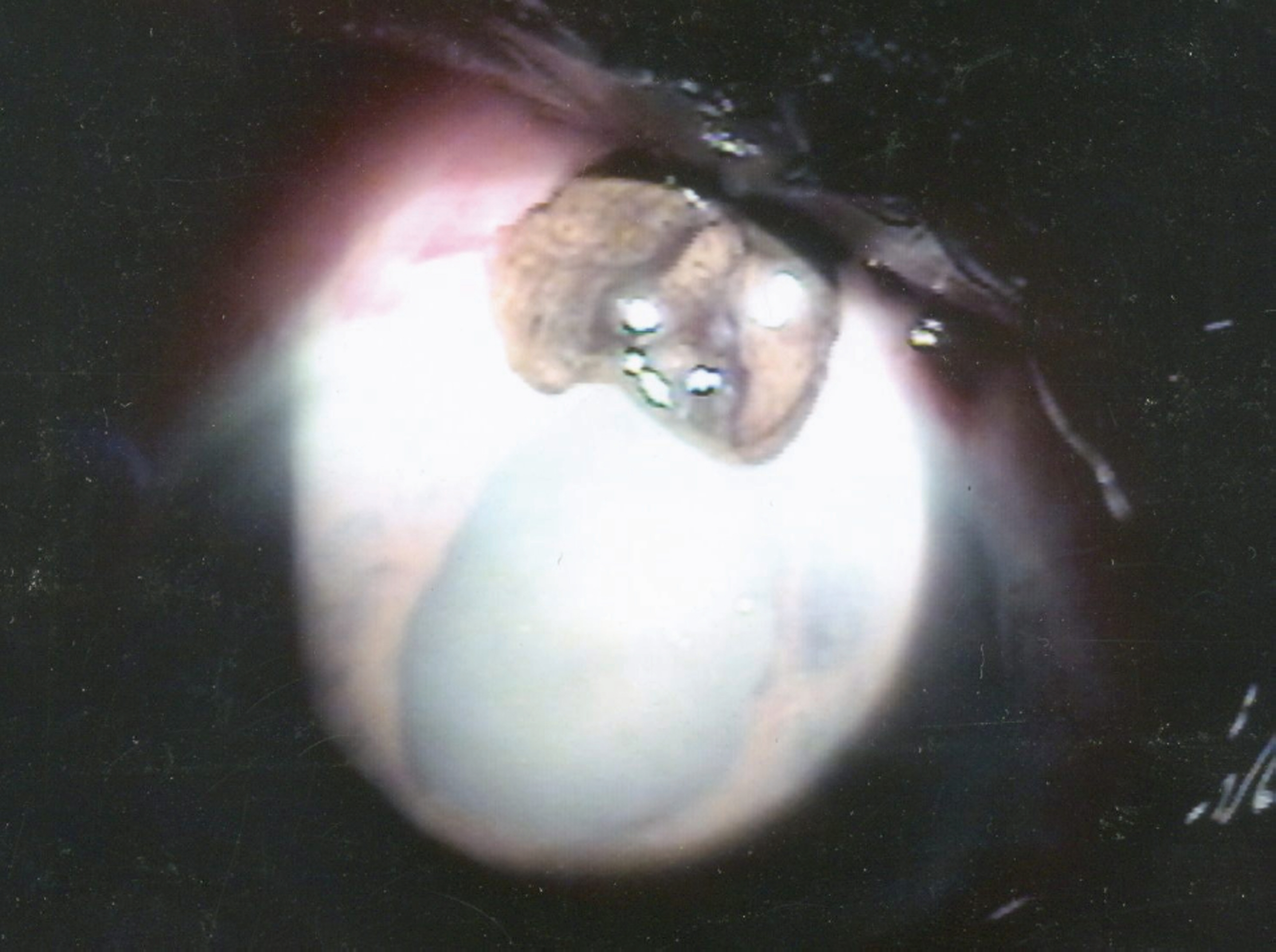
Figure 1. Preoperative anterior segment photograph of the right eye. It showed diffuse corneal edema and 5~6 mm full-thickness scleral laceration with protruded uveal tissue.

Figure 2. Postoperative anterior segment photograph. It showed autologous Tenon’s capsule patch graft (5×4 mm2), which was covered by a conjunctival flap at the superior limbal area.

Figure 3. (A) Three weeks postoperative picture. It showed ingrowth of fibrovascular patch over the Tenon’s capsule graft. (B) Six months postoperative picture. It showed successfully survived autologous Tenon’s capsule graft, which was completely covered by the fibrovascular patch.

Figure 4. (A) Anterior segment photograph after autograft of Tenon’s capsule. It showed mature cataract. (B) Anterior segment photograph 3 months after phacoemulsification and IOL implantation in the bag.
Reference
-
References
1. Lee CO, Jong SH, Lee JJ. Autologous Simple Conjunctival Graft and Conjunctiva/Tenon Graft on Focal Scleromalacia. J Korean Ophthalmol Soc. 1997; 38:1737–41.2. Rhee HS, Kim MS, Kim JH. Scleral graft on necrotic scleritis following pterygium excision. J Korean Ophthalmol Soc. 1987; 28:565–9.3. Na YS, Joo MJ, Kim JH. Results of scleral Allografting on scleral necrosis following pterygium excision. J Korean Ophthalmol Soc. 2005; 46:402–9.4. Sangwan VS, Jain V, Gupta P. Structural and functional outcome of scleral patch graft. Eye. 2007; 21:930–5.
Article5. Ozcan AA, Bilgic E, Yagmur M. Ersöz TR. Surgical management of scleral defects. Cornea. 2005; 24:308–11.6. Wang B, Wang Y, Ding J. Repair of cornea-sclera defect by autogenous sclera graft from the same eye. Zhongguo Xiu Fu Chong Jian Wai Ke Za Zhi. 1997; 11:206–7.7. Esquenazi S. Autogenous lamellar scleral graft in the treatment of scleral melt after pterygium surgery. Graefes Arch Clin Exp Ophthalmol. 2007; 245:1869–71.
Article8. Kwak JY, Chang HK. Autogenous temporalis fascia grafting and conjunctival flap transposition in scleromalacia after pterygium excision. J Korean Ophthalmol Soc. 2004; 45:180–6.9. Koenig SB, Sanitato JJ, Kaufman HE. Long-Term Follow-up Study of Scleroplasty Using Autogenous Periosteum. Cornea. 1990; 9:139–43.
Article10. Mauriello JA Jr, Pokorny K. Use of split-thickness dermal grafts to repair corneal and scleral defects-a study of 10 patients. Br J Ophthalmol. 1993; 77:327–31.
Article11. Hill RA, Aminlari A, Sassini JW, Michalski M. Use of symblepharon ring for treatment of over filtration and leaking blebs after glaucoma filtration surgery. Ophthalmic Surg. 1990; 21:707–10.
- Full Text Links
-
- Actions
-
Cited
- CITED
-
- Close
- Share
-
- Similar articles
-
- The Therapeutic Efficacy of Exposed Orbital Implant (Medpor(R)) Using Tenon's Capsule
- Rotation Flap of Tenon's Capsule for Treating Hypotony
- Autologous Simple Conjunctival Graft and Conjunctiva/Tenon Graft on Focal Scleromalacia
- Clinical Efficacy of Dermis-Fat Graft vs. Posterior Tenon's Capsule Suturing in Anophthalmic Orbit
- Lamellar Corneal Graft Using Acellular Preserved Human Cornea for Perforated Anterior Scleral Staphyloma
