Radiation Induced Hand Necrosis of an Orthopaedic Surgeon Who Had Treated a Patient with Fluoroscopy-Guided Spine Injection
- Affiliations
-
- 1Department of Orthopedic Surgery, School of Medicine, Wonkwang University, Iksan, Korea.
- 2Department of Orthopedic Surgery, Wonkwang University Sanbon Hospital, Gunpo, Korea. castkim@daum.net
Abstract
- As the frequency of radiation exposure by fluoroscopy continues to increase in orthopaedic fields, the level of hazard for the orthopaedic surgeon increases at the same time. Exposure of the clinician's hand is highest during performance of surgery or procedures within the actual clinics. Studies on radiation exposure on thyroid, eye or whole body, or reports on radiation treatment of cancer or for dermal lesions occurring from therapeutic intervention on the body such as heart and liver, and studies on radioactive damage to hands derived from radioactive material handlers have been reported; however, no studies on radioactive damage to a clinician's hand have been reported. Therefore, we report on a case of chronic radiation dermatitis and necrosis of an orthopaedic surgeon's hand as well as its soft tissue defect.
Figure
-
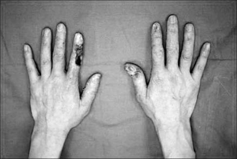
Figure 1 Radiation dermatitis developed on the hands of an orthopedic surgeon. Overall, both sides of the thumb and index finger showed redness, swelling, and atrophy of the nails. Necrosis measuring 6×4 cm2 was observed on the radial side of the left index finger.
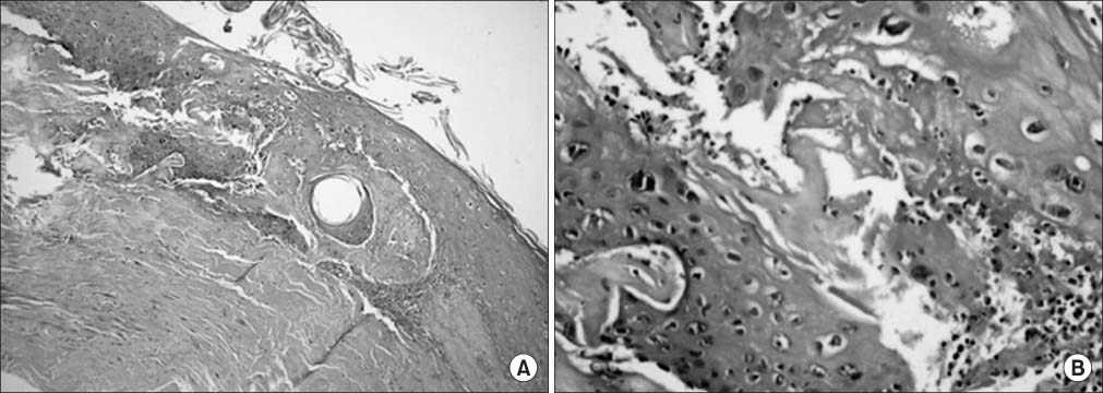
Figure 2 The biopsy shows features of chronic radiation dermatitis. (A) The biopsy shows epidermal atrophy, fibrotic dermis with degeneration, maturation destruction of membrane, and absence of hair follicles (H&E, ×40). (B) The microscopic pathologic findings of epidermis show severe inflammatory cell infiltration, atypia inflammation, and thickend collagen fibers (H&E, ×200).

Figure 3 An ulcerative lesion on the second finger was well treated using dorsalispedis free flap and split thickness skin-graft.
Reference
-
1. Maxon HR, Thomas SR, Saenger EL, Buncher CR, Kereiakes JG. Ionizing irradiation and the induction of clinically significant disease in the human thyroid gland. Am J Med. 1977; 63:967–978.
Article2. Merriam GR Jr, Focht EF. A clinical study of radiation cataracts and the relationship to dose. Am J Roentgenol Radium Ther Nucl Med. 1957; 77:759–785.3. Lee EW, Chun JM, Ahn BW, Park YW, Lee SY, Paik NC. A study of hand lesion exposed by radiation. J Korean Orthop Assoc. 1991; 26:841–846.
Article4. 1990 Recommendations of the International Commission on Radiological Protection. Ann ICRP. 1991; 21:1–201.5. Mehlman CT, DiPasquale TG. Radiation exposure to the orthopaedic surgical team during fluoroscopy: "how far away is far enough?". J Orthop Trauma. 1997; 11:392–398.
Article6. Noordeen MH, Shergill N, Twyman RS, Cobb JP, Briggs T. Hazard of ionizing radiation to trauma surgeons: reducing the risk. Injury. 1993; 24:562–564.
Article7. Arnstein PM, Richards AM, Putney R. The risk from radiation exposure during operative X-ray screening in hand surgery. J Hand Surg Br. 1994; 19:393–396.
Article8. Damilakis J, Koukourakis M, Hatjidakis A, Karabekios S, Gourtsoyiannis N. Radiation exposure to the hands of operators during angiographic procedures. Eur J Radiol. 1995; 21:72–75.
Article9. Goldsmith LA, Katz SI, Gilchrest BA, Paller AS, Leffell DJ, Wolff K. Fitzpatrick's dermatology in general medicine. 8th ed. New York: McGraw-Hill Medical;2012. p. 2893–2898.10. Elder DE, Elenitasas R, Johnson BL Jr, Murphy GF, Xu X. Lever's histopathology of the skin. 10th ed. Philadelphia: Lippincott Williams & Wilkins;2009. p. 348–352.
- Full Text Links
-
- Actions
-
Cited
- CITED
-
- Close
- Share
-
- Similar articles
-
- Ultrasound-Guided Intervention in Lumbar Spine
- Ultrasound-guided interventions for spinal pain
- Ultrasound-Guided Intervention in Cervical Spine
- Radiation exposure and its reduction in the fluoroscopic examination and fluoroscopy-guided interventional radiology
- Radiation Exposure to the Hand of a Spinal Interventionalist during Fluoroscopically Guided Procedures
