Simple Subperiosteal Hematoma with a Periosteal Reaction Mimicking a Malignancy
- Affiliations
-
- 1Department of Orthopedic Surgery, Chungnam National University School of Medicine, Daejeon, Korea. hyunsd@cnu.ac.kr
- 2Department of Orthopedic Surgery, Dong-A University School of Medicine, Busan, Korea.
Abstract
- A benign periosteal reaction, which can occur after trauma or stress, has a solid and uninterrupted appearance on radiography. In contrast, an aggressive periosteal reaction, which may indicate a malignancy, appears as a Codman's triangle or with a spiculated and sunburst pattern. In the present case, an 11-year-old boy with a previous injury to the distal radial growth plate presented with diffuse osteolysis on the distal radial metaphysis and decreased opacity of the lateral side cortex on plain radiograph. A Codman's triangle-like lesion was seen on the lateral side of the distal radius, and a few spicules were observed on the medial side of the distal radius. A T2-weighted coronal magnetic resonance image revealed a mass that had stripped the periosteum; the mass had heterogeneous signal intensity and a fl uid-fluid level on axial views. The margins of the mass were unclear, but enhanced. Suspecting a primary malignancy, we performed a biopsy. The pathology revealed that the mass was a simple hematoma.
Keyword
MeSH Terms
Figure
-

Figure 1 (A, B) A radiopaque shadow of the mass border was present on anteroposterior and lateral simple wrist radiographs, and diffuse osteolysis was seen at the distal radial metaphysis. The cortical shadow decreased on the lateral side of the metaphysis. A Codman's triangle-like lesion was seen on the lateral side of the distal radius, and a few spicules were observed on the medial cortex. A Salter-Harris classification I growth plate injury was found on the palmar side of the distal radius on a lateral radiograph, and a linear shadow of periosteal new bone formation was visible. (C, D) Coronal images showed an inhomogeneous high signal-intensity mass. The mass margins were ambiguous and enhanced. The mass was bulging, but the bone marrow showed normal signal intensity.
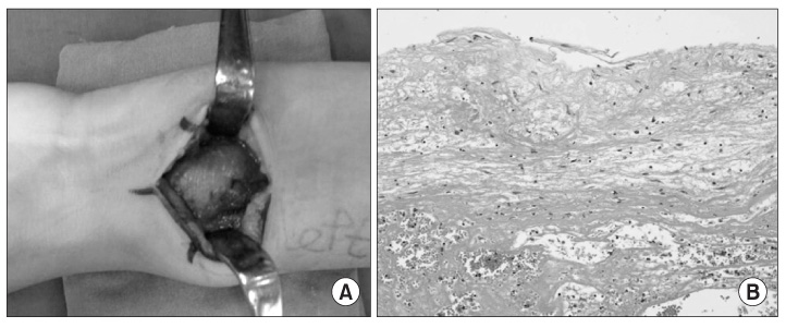
Figure 2 (A) The mass was located on the periosteum. The margin was clear, and the pronator quadratus was compressed. (B) Diffuse hemorrhage with fibrinoid degeneration and acute inflammation was shown at permanent biopsy (H&E stain, ×200).

Figure 3 (A, B) The spiculated pattern of the periosteal reaction and Codman's triangle-like lesion were not significant on follow-up radiographs taken 10 weeks later, but a double cortical shadow was present. The patient did not complain of any symptoms.
Reference
-
1. Miller TT. Bone tumors and tumorlike conditions: analysis with conventional radiography. Radiology. 2008. 246:662–674.
Article2. Ragsdale BD, Madewell JE, Sweet DE. Radiologic and pathologic analysis of solitary bone lesions. Part II: periosteal reactions. Radiol Clin North Am. 1981. 19:749–783.3. Koskinen SK, Mattila KT, Alanen AM, Aro HT. Stress fracture of the ulnar diaphysis in a recreational golfer. Clin J Sport Med. 1997. 7:63–65.
Article4. Ward WG Sr, Sekiya JK, Pope TL Jr. Traumatic ossifying periostitis of the ulna masquerading as a malignancy in a football player. A case report and literature review. Am J Sports Med. 1996. 24:852–856.5. Song KS, Kim HG, Park BM, Kim JM, Jung SH, Yang BS. Post traumatic osteolysis of the pubic bone simulating malignancy or osteomyelitis - a case report -. J Korean Bone Joint Tumor Soc. 2007. 13:180–184.6. Jahng J, Kim YH, Lee KS. Tuberculosis of the lower lumbar spine with an atypical radiological presentation -a case mimicking a malignancy-. Asian Spine J. 2007. 1:102–105.7. Shopfner CE. Periosteal bone growth in normal infants. A preliminary report. Am J Roentgenol Radium Ther Nucl Med. 1966. 97:154–163.8. Wenaden AE, Szyszko TA, Saifuddin A. Imaging of periosteal reactions associated with focal lesions of bone. Clin Radiol. 2005. 60:439–456.
Article9. Ishibashi Y, Okamura Y, Otsuka H, Nishizawa K, Sasaki T, Toh S. Comparison of scintigraphy and magnetic resonance imaging for stress injuries of bone. Clin J Sport Med. 2002. 12:79–84.
Article10. Resnick D. Diagnosis of bone and joint disorders. 2002. 4th ed. Philadelphia: WB Saunders;2346–2373.
- Full Text Links
-
- Actions
-
Cited
- CITED
-
- Close
- Share
-
- Similar articles
-
- Periosteal Reaction of Osteomyelitis: MRI Findings Compared with Plain Radiographs
- Exophthalmos Caused by Subperiosteal Orbital Hematoma
- Subperiosteal hematoma of the orbit associated with sinusitis
- A Case of Subperiosteal Orbital Hematoma in Patient with Ethmoid and Maxillary Sinusitis
- A Case of Subperiosteal Orbital Hematoma Associated with von Willebrand Disease
