Anatomic Characteristics of the Distal Femur and the Proximal Tibia in Koreans Undergoing Total Knee Arthroplasty
- Affiliations
-
- 1Center for Joint Disease, Chonnam National University Hwasun Hospital, Hwasun, Korea. eksong@jnu.ac.kr
- 2Department of Orthopaedic Surgery, Chonnam National University Hospital, Gwangju, Korea.
Abstract
- PURPOSE
In this study, we examined the anatomic characteristics of the distal femur and the proximal tibia to determine if there were significant differences between male and female patients.
MATERIALS AND METHODS
Seventy six patients (50 of female, 26 of male) who were treated by total knee arthroplasty using ROBODOC(R), were included. The anteroposterior and mediolateral length of the distal femur and proximal tibia were checked using the ORTHODOC program.
RESULTS
The mean sizes of the distal femur and proximal tibia of female knees were slightly smaller than those of male knees. Statistically significant differences were observed between the two groups (p<0.05 for all). On the other hand, the anteroposterior and mediolateral ratios of distal femur and proximal tibia revealed no differences between female and male knees (p>0.05 for all).
CONCLUSION
The anteroposterior to mediolateral ratio was similar in the distal femur and proximal tibia of Korean females.
Figure
-

Figure 1 Measurement of the anteroposterior length of lateral femoral condyle, length of line AB and anteroposterior distance from anterior diaphyseal cortex to most posterior point of lateral femoral condyle.
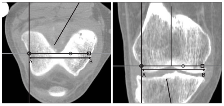
Figure 2 Measurement of the mediolateral length of Box, length of line AB and mediolateral distance between most prominent points mediolaterally at the level of distal femoral cutting.
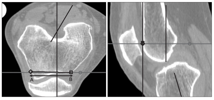
Figure 3 Measurement of the mediolateral length of anterior flange, length of line AB and mediolateral distance at the junction of planned anterior femoral and chamber cut.
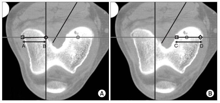
Figure 4 Measurement of the mediolateral length of lateral femoral condyle and mediolateral length of medial femoral condyle. (A) Length of line AB and mediolateral distance of lateral femoral condyle at the planned posterior femoral cut. (B) Length of line CD and mediolateral distance of medial femoral condyle at the planned posterior femoral cut.

Figure 5 Measurement of the anterior height of lateral femoral condyle and anterior height of medial femoral condyle. (A) Length of line AB and anterior height of lateral femoral condyle at the planned anterior femoral cut. (B) Length of line CD and anterior height of medial femoral condyle at the planned anterior femoral cut.
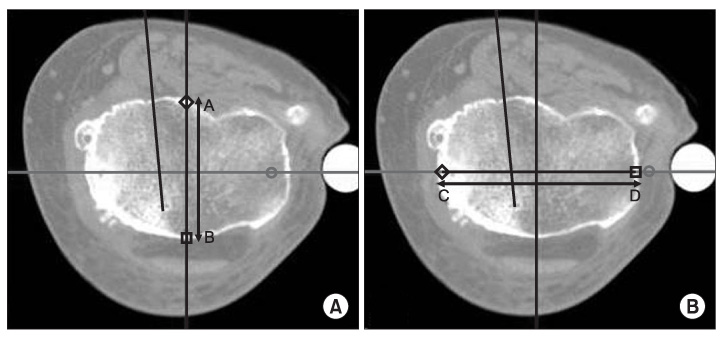
Figure 6 Measurement of the anterior height of middle of proximal tibia and mediolateral length of proximal tibia. (A) Length of line AB and anterior height of proximal tibia at the middle of longest mediolateral length. (B) Length of line CD and longest mediolateral length of proximal tibia parallel to the transepicondylar axis of distal femur at the planned anterior femoral cut.
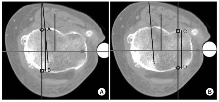
Figure 7 Measurement of the anterior height of lateral and medial condyle of proximal tibia. (A) Length of line AB and anterior height of lateral tibial condyle at the planned tibial cut. (B) Length of line CD and anterior height of medial tibial condyle at the planned tibial cut.

Figure 8 Measurement of the distance from the anterior height of lateral and medial condyle to the anterior height of middle of proximal tibia. (A) Length of line AB and distance from the line of CL to the line of AP at the planned tibial cut. (B) Length of line CD and distance from the line of CM to the line of AP at the planned tibial cut. CL, lateral to center distance; AP, anteroposterior; CM, medial to center distance.
Reference
-
1. Hootman JM, Sniezek JE, Helmick CG. Women and arthritis: burden, impact and prevention programs. J Womens Health Gend Based Med. 2002. 11:407–416.2. Kwak DS, Surendran S, Pengatteeri YH, et al. Morphometry of the proximal tibia to design the tibial component of total knee arthroplasty for the Korean population. Knee. 2007. 14:295–300.
Article3. Kane RL, Saleh KJ, Wilt TJ, Bershadsky B. The functional outcomes of total knee arthroplasty. J Bone Joint Surg Am. 2005. 87:1719–1724.
Article4. Roberts VI, Esler CN, Harper WM. A 15-year follow-up study of 4606 primary total knee replacements. J Bone Joint Surg Br. 2007. 89:1452–1456.
Article5. Vaidya SV, Ranawat CS, Aroojis A, Laud NS. Anthropometric measurements to design total knee prostheses for the Indian population. J Arthroplasty. 2000. 15:79–85.
Article6. Bengs BC, Scott RD. The effect of patellar thickness on intraoperative knee flexion and patellar tracking in total knee arthroplasty. J Arthroplasty. 2006. 21:650–655.
Article7. Parsley BS, Bertolusso R, Harrington M, Brekke A, Noble PC. Influence of gender on age of treatment with TKA and functional outcome. Clin Orthop Relat Res. 2010. 468:1759–1764.
Article8. Vincent KR, Vincent HK, Lee LW, Alfano AP. Outcomes in total knee arthroplasty patients after inpatient rehabilitation: influence of age and gender. Am J Phys Med Rehabil. 2006. 85:482–489.9. Conley S, Rosenberg A, Crowninshield R. The female knee: anatomic variations. J Am Acad Orthop Surg. 2007. 15:Suppl 1. S31–S36.
Article10. Fehring TK, Odum SM, Hughes J, Springer BD, Beaver WB Jr. Differences between the sexes in the anatomy of the anterior condyle of the knee. J Bone Joint Surg Am. 2009. 91:2335–2341.
Article11. Hitt K, Shurman JR 2nd, Greene K, et al. Anthropometric measurements of the human knee: correlation to the sizing of current knee arthroplasty systems. J Bone Joint Surg Am. 2003. 85:Suppl 4. 115–122.12. Dalury DF, Mason JB, Murphy JA, Adams MJ. Analysis of the outcome in male and female patients using a unisex total knee replacement system. J Bone Joint Surg Br. 2009. 91:357–360.
Article13. MacDonald SJ, Charron KD, Bourne RB, Naudie DD, McCalden RW, Rorabeck CH. The John Insall Award: gender-specific total knee replacement: prospectively collected clinical outcomes. Clin Orthop Relat Res. 2008. 466:2612–2616.
Article14. Merchant AC, Arendt EA, Dye SF, et al. The female knee: anatomic variations and the female-specific total knee design. Clin Orthop Relat Res. 2008. 466:3059–3065.
Article15. Greene KA. Gender-specific design in total knee arthroplasty. J Arthroplasty. 2007. 22(7):Suppl 3. 27–31.
Article16. Clarke HD, Hentz JG. Restoration of femoral anatomy in TKA with unisex and gender-specific components. Clin Orthop Relat Res. 2008. 466:2711–2716.
Article17. Emerson RH Jr, Martinez J. Men versus women: does size matter in total knee arthroplasty? Clin Orthop Relat Res. 2008. 466:2706–2710.18. Kim YH, Choi Y, Kim JS. Comparison of standard and gender-specific posterior-cruciate-retaining high-flexion total knee replacements: a prospective, randomised study. J Bone Joint Surg Br. 2010. 92:639–645.19. Lonner JH, Jasko JG, Thomas BS. Anthropomorphic differences between the distal femora of men and women. Clin Orthop Relat Res. 2008. 466:2724–2729.
Article20. Poilvache PL, Insall JN, Scuderi GR, Font-Rodriguez DE. Rotational landmarks and sizing of the distal femur in total knee arthroplasty. Clin Orthop Relat Res. 1996. (331):35–46.
Article21. Chin KR, Dalury DF, Zurakowski D, Scott RD. Intraoperative measurements of male and female distal femurs during primary total knee arthroplasty. J Knee Surg. 2002. 15:213–217.
- Full Text Links
-
- Actions
-
Cited
- CITED
-
- Close
- Share
-
- Similar articles
-
- The Bone Mineral Density of the Proximal Tibia, Lumbar Spine and Proximal Femur and Its Correlation with the Alignment of the Lower Extremity in Knee Osteoarthritic Patients
- Functional Evaluation of Tumor Prosthetic Arthroplasty for the Malignant Bone Tumor around the Knee: Interim Report
- The Study of Anatomical Measurement of Distal Femur and Compatibility of Femoral Prosthesis in Total Knee Arthroplasty
- Cement Mantle Thickness at the Bone Cement Interface in Total Knee Arthroplasty: Comparison of PS150 RP and LPS-Flex Knee Implants
- Indirect Measurement of Joint Space Distance using Ladmarks in TKRA
