Midterm Clinical and Radiological Results after Microfracture in Osteoarthritic Knees
- Affiliations
-
- 1Department of Orthopedic Surgery, School of Medicine, Kyung Hee University, Seoul, Korea. bdkyung@khmc.or.kr
Abstract
-
PURPOSE: To evaluate midterm results after microfracture in osteoarthritic knees.
MATERIALS AND METHODS
Between October 1997 and April 2006, 67 osteoarthritic knees, with minimum 4-year follow-up, underwent microfracture. Baumgaertner scores were evaluated to determine clinical results. Radiological results were assessed based on joint space widening and improvement of mechanical axis deviation. Joint space widening was calculated by comparing the preoperative joint space with the final follow-up joint space. Varus deformity was evaluated on orthoroentgenogram and recorded as a percentile of the point at which the mechanical axis intersected a line extending from the center of the knee to medial border of the medial tibial condyle. The figure was expressed as MA%.
RESULTS
The average Baumgaertner score at final follow-up was 7.0. The average joint space changed from 2.74 mm to 4.22 mm on AP radiographs and from 1.91 mm to 3.85 mm on lateral radiographs. Average MA% was 57.5% preoperatively and 45.8% at final follow-up. Clinical and radiological improvements were maintained in most cases followed for more than 4 years. Four patients had total knee arthroplasty after an average of 4.1 years.
CONCLUSION
We noted pain relief, joint space widening, and improvement of mechanical axis after microfracture for degenerative arthritis of the knee. Maintenance of clinical and radiological improvement was observed at midterm follow-up.
Keyword
MeSH Terms
Figure
-
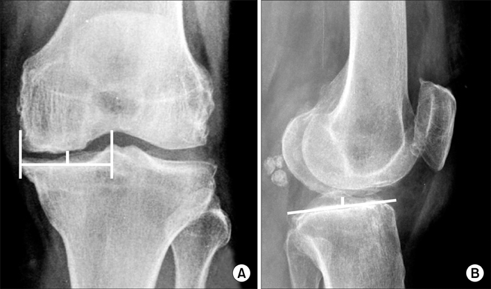
Fig. 1 (A) Joint space on the AP radiograph was measured by the distance between the medial femoral condyle and the midpoint from medial tibial spine to medial border of the medial tibial condyle. (B) Joint space on the lateral radiograph was measured by the nearest of distance between medial femoral condyle and medial tibial plateau.

Fig. 2 The extent of varus deformity shown on preoperative orthoroentgenogram was evaluated by MA% [(b/a)×100]. It shows a medial deviation of the mechanical axis with joint space narrowing of medial compartment: (a) is the distance from the center of the knee to the medial border of the medial tibial condyle and (b) is the distance from the center of the knee to the point at which the mechanical axis intersects the knee joint line.
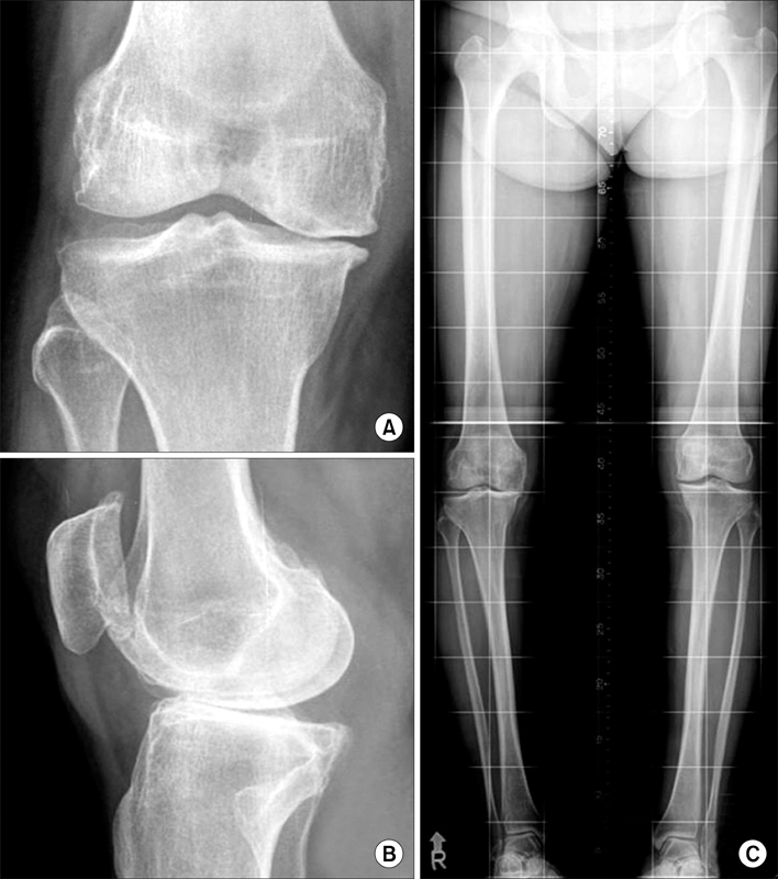
Fig. 3 Preoperative radiographs of fifty five-year-old woman with symptomatic degenerative osteoarthritis on right knee. (A) AP gap was 2.21 mm on anterior-posterior view of the knee. (B) Lateral gap was 1.19 mm on lateral view of the knee. (C) MA% was 99.8% on orthoroentgenogram.
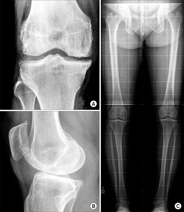
Fig. 4 Radiographs 4 years after microfracture of fifty five-year-old woman with symptomatic degenerative osteoarthritis on right knee. (A) AP gap was 4.40 mm on anteriorposterior view of the knee. (B) Lateral gap was 2.48 mm on lateral view of the knee. (C) MA% was 52.1% on orthoroentgenogram.
Reference
-
1. Bae DK, Ko BW, Kim SK. Results of microfracture surgery in osteoarthritic knee. J Korean Orthop Assoc. 2001. 36:555–560.
Article2. Bae DK, Yim CM, Kim JM, Park YK. Microfracture surgery for cartilage regeneration in degenerative arthritis of the knee. J Korean Orthop Assoc. 2000. 35:231–238.
Article3. Bae DK, Yoon KH, Song SJ. Cartilage healing after microfracture in osteoarthritic knees. Arthroscopy. 2006. 22:367–374.
Article4. Bassett CA. Bibliography of bone formation. Transplant Bull. 1962. 29:104–109.5. Baumgaertner MR, Cannon WD Jr, Vittori JM, Schmidt ES, Maurer RC. Arthroscopic debridement of the arthritic knee. Clin Orthop Relat Res. 1990. 253:197–202.
Article6. Blevins FT, Steadman JR, Rodrigo JJ, Silliman J. Treatment of articular cartilage defects in athletes: an analysis of functional outcome and lesion appearance. Orthopedics. 1998. 21:761–767.
Article7. Dandy DJ. Arthroscopic debridement of the knee for osteoarthritis. J Bone Joint Surg Br. 1991. 73:877–878.
Article8. Friedman MJ, Berasi CC, Fox JM, Del Piazzo W, Snyder SJ, Ferkel RD. Preliminary results with abrasion arthroplastyin the osteoarthritic knee. Clin Orthop Relat Res. 1984. 182:200–205.9. Gobbi A, Nunag P, Malinowski K. Treatment of full thickness chondral lesions of the knee with microfracture in a group of athletes. Knee Surg Sports Traumatol Arthrosc. 2005. 13:213–221.
Article10. Johnson LL. Arthroscopic abrasion arthroplasty historical and pathologic perspective: present status. Arthroscopy. 1986. 2:54–69.
Article11. Kellgren JH, Lawrence JS. Radiological assessment of osteo-arthrosis. Ann Rheum Dis. 1957. 16:494–502.
Article12. Knutsen G, Engebretsen L, Ludvigsen TC, et al. Autologous chondrocyte implantation compared with microfracture in the knee. A randomized trial. J Bone Joint Surg Am. 2004. 86:455–464.13. Kuo AC, Rodrigo JJ, Reddi AH, Curtiss S, Grotkopp E, Chiu M. Microfracture and bone morphogenetic protein 7 (BMP-7) synergistically stimulate articular cartilage repair. Osteoarthritis Cartilage. 2006. 14:1126–1135.
Article14. Marder RA, Hopkins G Jr, Timmerman LA. Arthroscopic microfracture of chondral defects of the knee: a comparison of two postoperative treatments. Arthroscopy. 2005. 21:152–158.
Article15. Miller BS, Steadman JR, Briggs KK, Rodrigo JJ, Rodkey WG. Patient satisfaction and outcome after microfracture of the degenerative knee. J Knee Surg. 2004. 17:13–17.
Article16. Mithoefer K, Williams RJ 3rd, Warren RF, et al. The microfracture technique for the treatment of articular cartilage lesions in the knee. A prospective cohort study. J Bone Joint Surg Am. 2005. 87:1911–1920.17. Ogilvie-Harris DJ, Fitsialos DP. Arthroscopic management of the degenerative knee. Arthroscopy. 1991. 7:151–157.
Article18. Passler HH. Microfracture for treatment of cartilage defects. Zentralbl Chir. 2000. 125:500–504.19. Qui YS, Shahgaldi BR, Revel WJ, Heatley FW. Observation of subchondral plate advancement during osteochondral repair: a histomorphometric and mechanical study in the rabbit femoral condyle. Osteoarthritis Cartilage. 2003. 11:810–820.20. Rodrigo J, Steadman JR, Syftestad G, Benton H, Silliman J. Effects of human knee synovial fluid on chondrogenesis in vitro. Am J Knee Surg. 1995. 8:124–129.21. Steadman JR, Briggs KK, Rodrigo JJ, Kocher MS, Gill TJ, Rodkey WG. Outcomes of microfracture for traumatic chondral defects of the knee: average 11-year follow-up. Arthroscopy. 2003. 19:477–484.
Article22. Steadman JR, Miller BS, Karas SG, Schlegel TF, Briggs KK, Hawkins RJ. The microfracture technique in the treatment of full-thickness chondral lesions of the knee in National Football League players. J Knee Surg. 2003. 16:83–86.23. Steadman JR, Rodkey WG, Briggs KK, Rodrigo JJ. The microfracture technic in the management of complete cartilage defects in the knee joint. Orthopade. 1999. 28:26–32.24. Sterett WI, Steadman JR. Chondral resurfacing and high tibial osteotomy in varus knee. Am J Sports Med. 2004. 32:1243–1249.25. Urist MR. The repair of articular surfaces following arthroplasty of the hip. Clin Orthop. 1958. 12:209–229.26. Woo SL, Kwan MK, Lee TQ, Field FP, Kleiner JB, Coutts RD. Perichondral autograft for articular cartilage. Shear modulus of neocartilage studied in rabbits. Acta Orthop Scand. 1987. 58:510–515.
- Full Text Links
-
- Actions
-
Cited
- CITED
-
- Close
- Share
-
- Similar articles
-
- Results of Microfracture Surgery in Osteoarthritic Knee
- Results of Microfracture in the Osteoarthritic Knee with Focal Full-Thickness Articular Cartilage Defects and Concomitant Medial Meniscal Tears
- Immunohistochemical Staining for Type II Collagen in Regenerated Cartilage after Microfracture Surgery
- Quantitative Analysis of Type II Collagen with Western Blotting in Microfracture Surgery
- Autologous Chondrocyte Implantation as a Secondary Procedure after Failed Microfracture for Osteochondral Lesion of Talus
