365 nm LED laser treatment on beagle for gingival whitening without gum dermabrasion
- Affiliations
-
- 1Department of Oral and Maxillofacial Surgery, Clinical Trial Center, Seoul National University Dental Hospital, Seoul, Republic of Korea. leejongh@snu.ac.kr
- 2College of information and Communication Engineering, Sungkyunkwan University, Suwon, Republic of Korea.
- 3Department of Oral and Maxillofacial Surgery, Korea University Medical Center, Guro Hospital, Seoul, Republic of Korea.
- 4Department of Oral and Maxillofacial Surgery, Dongtan Sacred Heart Hospital, Hallym University Medical Center, Suwon, Republic of Korea.
- 5Department of Prosthodontics, Seoul National University Dental Hospital, Seoul, Republic of Korea.
- 6Dental Research Institute, School of Dentistry, Seoul National University, Seoul, Republic of Korea.
- KMID: 2328893
- DOI: http://doi.org/10.14368/jdras.2016.32.2.117
Abstract
- PURPOSE
Gingival whitening is one of dental treatment purposes which is close to treating aesthetic disorders. Initial gingival whitening treatment was done by dermabrasion using a high power Diode Laser. However, this treatment method cannot be free from any infection or pain after the treatment. Therefore, we have decided to progress gingival whitening treatment using a low power LED laser.
MATERIALS AND METHODS
The laser was irradiated on pork meat then the safety of output power, temperature change and skin denaturalization was measured. Bison 365 nm LED laser was irradiated on oral mucosal pigment of a 15 - 20 kg beagle for 15 min for 1 - 2 weeks, one or two times each. Any pigment loss was checked through Hematoxyline-Eosin staining.
RESULTS
The melanin pigments at the area of 365 nm LED Laser irradiation were decreased.
CONCLUSION
The 365 nm LED Laser proposed in this study is considered to compensate the bleaching effect achieved by either using Diode laser or surgical methods.
Keyword
MeSH Terms
Figure
-
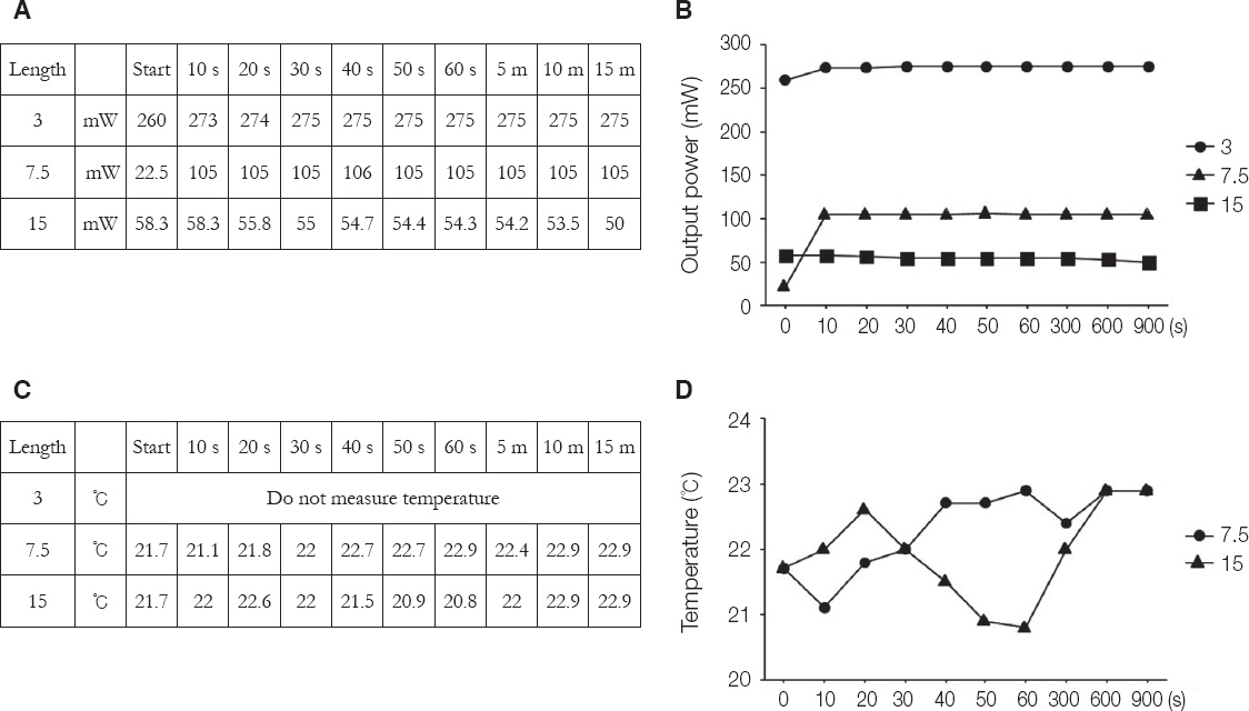
Fig. 1 Bison 365 nm LED laser output power & Temperature. (A, B) Change in output power when irradiated 365 nm laser on pork, (C, D) Measurement of change in temperature when irradiated 365 nm laser. (C) 3°C was not measured because it exceeded the 100 mW.
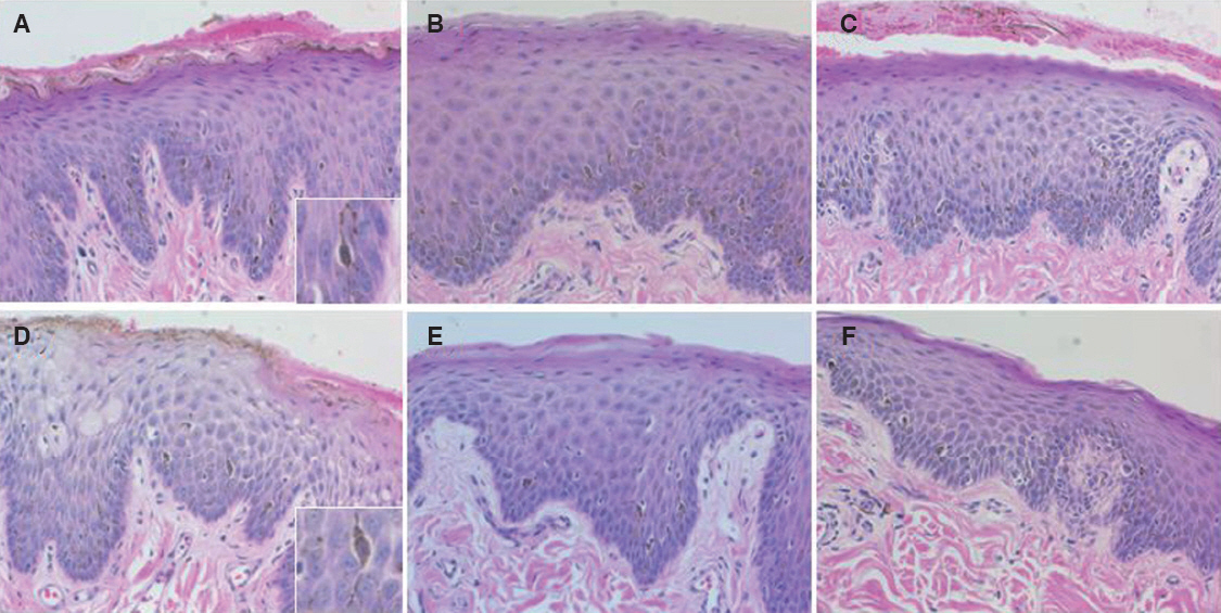
Fig. 2 Melanin pigment H/E staining. (A, D) 1 weeks and 2 weeks control groups. (B, E) It was melanin pigment that stained H/E in 1 weeks 1-time and 2 weeks 1-time irradiation. (C, F) It was melanin pigment that stained H/E in 1 weeks 2-time and 2 weeks 2-time irradiation.

Fig. 3 Melanin pigment counting statistical analysis. (A) Comparison of 1 weeks 1-time irradiation and 1 weeks 2-time (*, **P < 0.05). (B) Comparison of 2 weeks 1-time irradiation and 2 weeks 2-time (* P < 0.05). (C) Comparison of 1 weeks 1-time irradiation and 2 weeks 1-time irradiation (*, ** P < 0.05). (D) Comparison of 1 weeks 2-time irradiation and 2 weeks 2-time irradiation (* P < 0.01 and ** P < 0.05).
Reference
-
References
1. Yousuf A, Hossain M, Nakamura Y, Yamada Y, Kinoshita J, Matsumoto K. Removal of gingival melanin pigmentation with the semiconductor diode laser: a case report. J Clin Laser Med Surg. 2000; 18:263–6. PMID: 11572242.2. Patil KP, Joshi V, Waghmode V, Kanakdande V. Gingival depigmentation : a split mouth comparative study between scalpel and cryosurgery. Contemp Clin Dent. 2015; 6:S97–S101. DOI: 10.4103/0976-237X.152964. PMID: 25821386. PMCID: PMC4374330.3. Monteiro LS, Costa JA, da Câmara MI, Albuquerque R, Martins M, Pacheco JJ, Salazar F, Figueira F. Aesthetic depigmentation of gingival smoker’s melanosis using carbon dioxide lasers. Case Rep Dent. 2015; 2015:510589. DOI: 10.1155/2015/510589.4. Chandna S, Kedige SD. Evaluation of pain on use of electrosurgery and diode lasers in the management of gingival hyperpigmentation: a comparative study. J Indian Soc Periodontol. 2015; 19:49–55. DOI: 10.4103/0972-124X.145823. PMID: 25810593. PMCID: PMC4365157.5. Fekrazad R, Chiniforush N. One visit providing desirable smile by laser application. J Lasers Med Sci. 2014; 5:47–50. PMID: 25606339. PMCID: PMC4290524.6. Berk G, Atici K, Berk N. Treatment of gingival pigmentation with Er, Cr: YSGG Laser. J Oral Laser Applications. 2005; 5:249–53.7. Soliman MM, Al Thomali Y, Al Shammrani A, El Gazaerly H. The use of soft tissue diode laser in the treatment of oral hyper pigmentation. Int J Health Sci. 2014; 8:133–140. DOI: 10.12816/0006079.
- Full Text Links
-
- Actions
-
Cited
- CITED
-
- Close
- Share
-
- Similar articles
-
- Laser-Dermabrasion as a Surgical Treatment for Cutaneous Scars and Wrinkles
- Effectiveness of low-level laser therapy and chewing gum in reducing orthodontic pain: A randomized controlled trial
- A Case of Refractory Vitiligo That Was Treated with a Combination of Non-ablative 1550-nm Erbium:Glass Fractional Laser, Narrow-band UVB, and a Topical Agent
- Application of Teeth Whitening LED for Prevention of Dental Caries : Antimicrobial Photodynamic Therapy Approach
- Chemical Peeling and Laser Resurfacing
