Ultrasound Evaluation of Ulnar Neuropathy at the Elbow Caused by a Mass Lesion
- Affiliations
-
- 1Department of Neurology, Yeungnam University College of Medicine, Daegu, Korea. minsupark@ynu.ac.kr
- KMID: 2328821
- DOI: http://doi.org/10.14253/kjcn.2016.18.1.7
Abstract
- Ulnar neuropathy at the elbow (UNE) may seem easy to diagnose when the characteristic clinical manifestations are present, and electrodiagnostic studies have high sensitivity, although they are non-localizing in some cases and unable to reveal structural lesions. Ultrasonography is noninvasive and able to find the exact location of the lesion and visualize perineural structures. We present two cases of UNE in which we found hypoechoic mass lesions near medial epicondyle with ultrasonography and discuss its usefulness in diagnosis of UNE.
Keyword
Figure
-
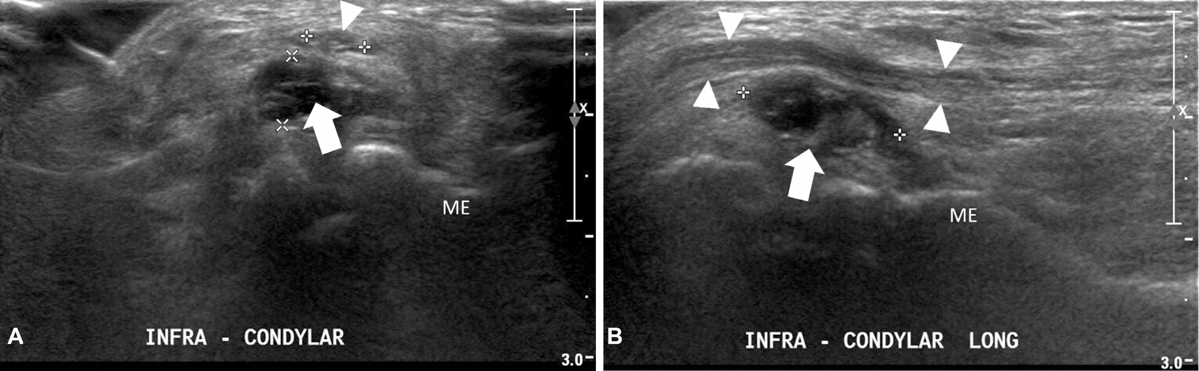
Figure 1. Nerve ultrasonography of case 1. Transverse (A) and longitudinal (B) views of the right elbow show ulnar nerve (arrowheads) and a hypoechogenic mass (arrows) compressing ulnar nerve below medial epicondyle. ME; medial epicondyle.
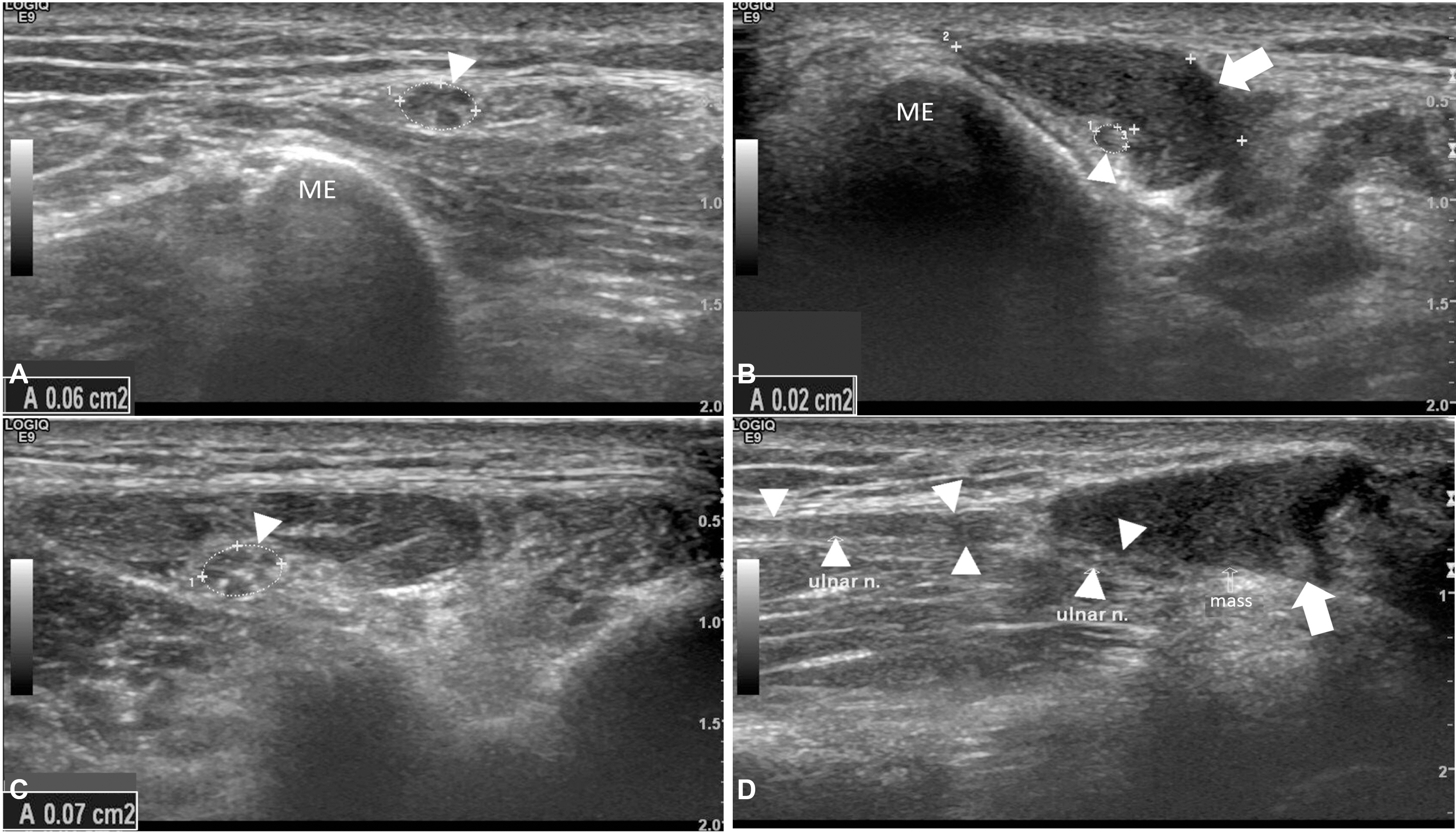
Figure 2. Nerve ultrasonography of case 2. Transverse view (A-C) tracing right ulnar nerve (arrowheads) reveals decreased cross sectional area (CSA) of the ulnar nerve in the level of the mass (arrow) (B) near the medial epicondyle and normal CSA in the proximal (A) and distal (C) parts. Longitudinal view (D) shows compression of the ulnar nerve (arrowheads) by the hypoechogenic mass (arrow). A; cross sectional area, ME; medial epicondyle.

Figure 3. Intraoperative photograph of case 2. The mass lesion seems rather enclosed by epineurium than isolated from the ulnar nerve as a cyst.
Reference
-
1.Beekman R., Visser LH., Verhagen WI. Ultrasonography in ulnar ne uropathy at the elbow: a critical review. Muscle Nerve. 2011. 43:627 -635.2.Omejec G., Žgur T., Podnar S. Diagnostic accuracy of ultrasonographic and nerve conduction studies in ulnar neuropathy at the elbow. Clin Neurophysiol. 2015. 126:1797–1804.
Article3.Ohana M., Moser T., Moussaouï A., Kremer S., Carlier RY., Liver-neaux P, et al. Current and future imaging of the peripheral nervous system. Diagn Interv Imaging. 2014. 95:17–26.
Article4.Stewart JD. The variable clinical manifestations of ulnar neuropathies at the elbow. J Neurol Neurosurg Psychiatry. 1987. 50:252–25. 8.
Article5.Campbell WW., Greenberg MK., Krendel DA., Pridgeon RM., Sitaram KP., Williams FH. The electrodiagnostic evaluation of patients with ulnar neuropathy at the elbow: literature review of the usefulness of nerve conduction studies and electromyography. Muscle Nerve. 1999. 22:S175–S205.6.Omejec G., Podnar S. Normative values for short-segment nerve conduction studies and ultrasonography of the ulnar nerve at the elbow. Muscle Nerve. 2015. 51:370–377.
Article7.van Veen KE., Wesstein M., van Kasteel V. Ultrasonography and electrodiagnostic studies in ulnar neuropathy: an examination of the sensitivity and specificity and the correlations between both diagnostic tools. J Clin Neurophysiol. 2015. 32:240–243.8.Beekman R., Schoemaker MC., Van Der Plas JP., Van Den Berg LH., Franssen H., Wokke JH, et al. Diagnostic value of high-resolution sonography in ulnar neuropathy at the elbow. Neurology. 2004. 62:767–773.
Article
- Full Text Links
-
- Actions
-
Cited
- CITED
-
- Close
- Share
-
- Similar articles
-
- Diagnosis of Ulnar Neuropathy Caused by Intraneural Ganglion at Elbow with Ultrasound
- High Ulnar Nerve Palsy by the Arcade of Struthers in the Elbow: Report of 2 Cases
- Subclinical Ulnar Neuropathy at the Elbow in Diabetic Patients
- Ultrasonographic Findings and Usefulness in Ulnar Neuropathy at the Elbow
- Tardy Ulnar Nerve Palsy by Neurofibroma
